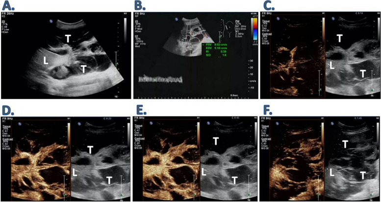
Fig. 2.
Ultrasound presentation of a patient with single massive primary hepatic angiosarcoma (congenital type). Patient 5. This figure illustrates a patient with a massive primary hepatic angiosarcoma located on the surface of the liver. The tumor exhibits a cystic and solid structure, growing downwards and compressing the right appendage. A and B Conventional ultrasound findings: The tumor surrounds the liver's surface, displaying uneven internal ultrasound and unclear boundaries. An interstitial septal pattern is present, with abundant level 3 blood flow signals visible within. The resistance index is 0.39; C Contrast-enhanced ultrasound performance: At 14 s, the interstitial septation within the lesion enhanced faster than the liver parenchyma; D The peak was reached in 22 s, with the interstitial septation within the lesion exhibiting slightly higher intensity than the liver parenchyma; E The cystic part within the tumor compartment remained unfilled at 45 s, and the strength of the interstitial septation equaled that of the liver parenchyma; F After 105 s, the intensity of the interstitial septa within the tumor gradually subsided. L: Liver; T: Tumor

