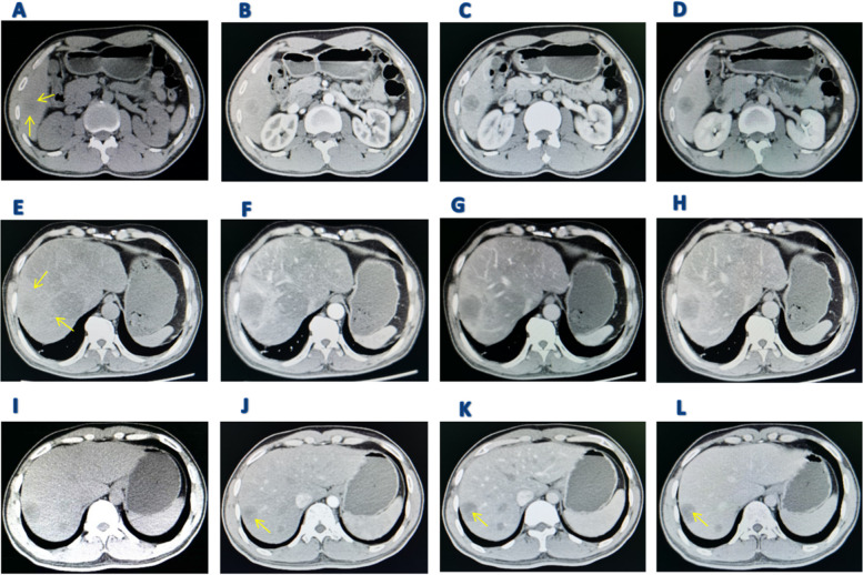
Fig. 3.
Computed tomography presentation of primary hepatic angiosarcoma. A Patient 6: Low-density shadows are evident during the plain scan, with clear boundaries (yellow arrows); B Mild circular enhancement around the arterial phase, along with several punctate nodular enhancements within the lesion, varying in intensity and mixed; C and D In the portal and delayed phases, peripheral enhancement of the lesion gradually diminishes, while internal nodular mixed signals persist; E Patient 7: During the plain scan, peripheral high-density shadows are visible, with low-density shadows at the center and unclear boundaries (yellow arrows); F In the arterial phase, the lesion's contour gradually sharpens as the surrounding ring strengthens; G and H In the portal and delayed phases, the lesion's enhancement subsides, but the interior remains unfilled and structurally disordered; I Patient 6: Low-density shadows are noticeable during the plain scan, featuring clear boundaries; J Mild peripheral enhancement during the arterial phase, featuring a "vascular sign" (yellow arrow); K and L In the portal and delayed phases, peripheral enhancement gradually diminishes, yet the "vascular sign" remains visible (yellow arrow)

