
FIG. 1.
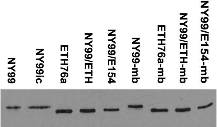
Western blot showing the differing mobilities of E proteins of WNV strains consistent with the presence of Asn-Tyr-Ser glycosylation motif or mutant Ser-Tyr-Ser at residues 154 to 156. Proteins were separated on a 5%/10% discontinuous sodium dodecyl sulfate-polyacrylamide gel electrophoresis gel, electrophoretically transferred to 0.2-μm nitrocellulose, and detected using WNV-specific monoclonal antibody 7H2. The suffix “mb” indicates strains isolated from the brain of a mouse that died following peripheral inoculation with the respective parent strain.