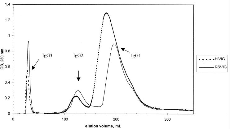
FIG. 1.
Separation of antibody subclasses from polyclonal immune globulin preparations by FPLC. RSVIG and HIVIG were run under identical conditions, and the graphs indicating the protein concentrations of the elution-fractions are superimposed. IgG3 protein was present in the initial flowthrough, as indicated. Upon application of a gradually diminishing pH gradient, IgG2 was eluted, followed and partially overlapped by IgG1. OD, optical density.

