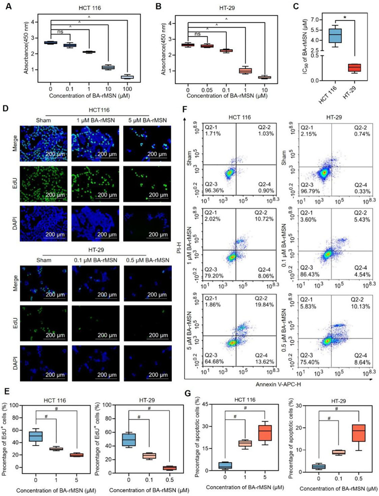
Fig. 2.
BA-rMSNs inhibit proliferation and induce cell death. (A) HCT 116 cells and (B) HT-29 cells were treated with PBS or different dosages of BA-rMSNs for 48 h. The results of the CCK8 assay revealed that BA-rMSNs significantly inhibited the viability of these cells. (C) The IC50 values indicated that, compared with HCT 116 cells, HT-29 cells were more sensitive to BA-rMSNs. Representative fluorescence microscopy images (D) and quantitative analysis of EdU (E) confirmed that BA-rMSNs significantly inhibited proliferation. (F) Annexin V-APC/PI staining revealed that BA-rMSNs increased the percentages of Annexin V-APC+/PI- (early apoptotic) cells and Annexin V-APC+/PI+ (late apoptotic) cells. (G) Quantification of the percentage of apoptotic HCT 116 and HT-29 cells. and E-G. ns indicates no significance, * indicates a P value lower than 0.05, # indicates a P value lower than 0.0167, and ^ indicates a P value lower than 0.0125, determined via the Mann‒Whitney U test. IC50: half-maximal inhibitory concentration

