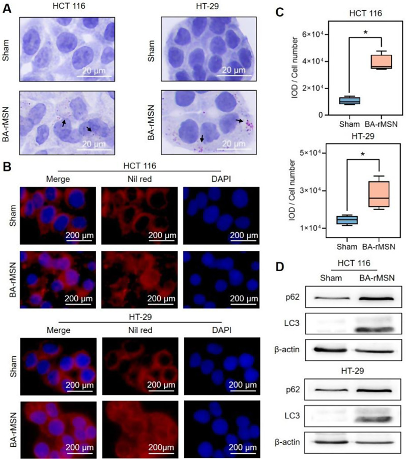
Fig. 3.
BA-rMSNs blocked lipophagy. (A) Oil red O staining and (B) Nile red staining demonstrated that BA-rMSNs increased the accumulation of lipid droplets. (C) Quantitative analysis of Nile red staining results in HCT 116 and HT-29 cells revealed an increase in lipid droplet accumulation. (D) Western blot analysis revealed that BA-rMSNs increased the levels of p62 and LC3, indicating the inhibition of lipophagy. N = 4 for B. * indicates a P value lower than 0.05, which was determined by the Mann‒Whitney U test. IOD: Integrated optical density

