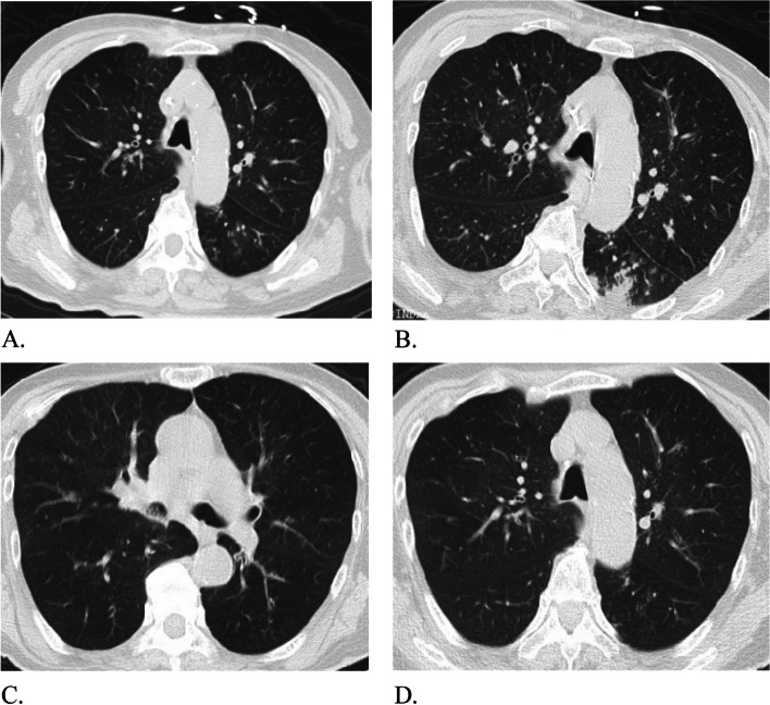
Fig. 2.
The changes of lung CT images. A The CT images on the 3rd day of mechanical ventilation. These images showed that there were micro-nodules distributed along the bronchovascular bundle in both lungs, a number of buds like changes, multiple signet ring signs and double track signs were seen locally, and the walls were thickened with patchy fuzzy shadow around. B The lung CT images on the 6th day of mechanical ventilation. Compared with the CT images on the 3rd day, the inflammation in the left lower lobe of the lung was aggravated. C and D The lung CT images after the withdraw of mechanical ventilation. Compared with the 6th day lung CT images, the inflammation of the left lower lung lobe was alleviated. At the same time, patchy high-density shadows were found in the middle bronchus and lower lobe bronchus of the right lung, which was considered to be sputum retention in the cavity

