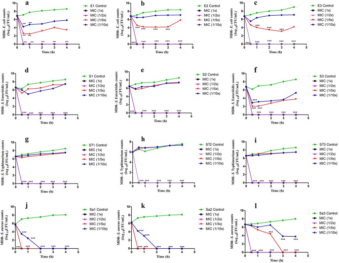
Fig. 2.
In vitro dose- and time-dependent photocatalytic disinfection kinetics of MDR bacterial strains treated with AgNPs. Images (a, b, c) denote the photocatalytic disinfection kinetics of MDR strains of EAEC (E1, E2, E3), images (d, e, f) represent photocatalytic disinfection kinetics of MDR strains of S. Enteritidis (S1, S2, S3), images (g, h, i) denote photocatalytic disinfection kinetics of MDR strains of S. Typhimurium (ST1, ST2, ST3) and images (j, k, l) denote photocatalytic disinfection kinetics of MDR strains of MRSA (Sa1, Sa2, Sa3) when treated with different concentrations of AgNPs (MIC 1X, 1/2X, 1/5X, 1/10X) under LED light

