Abstract
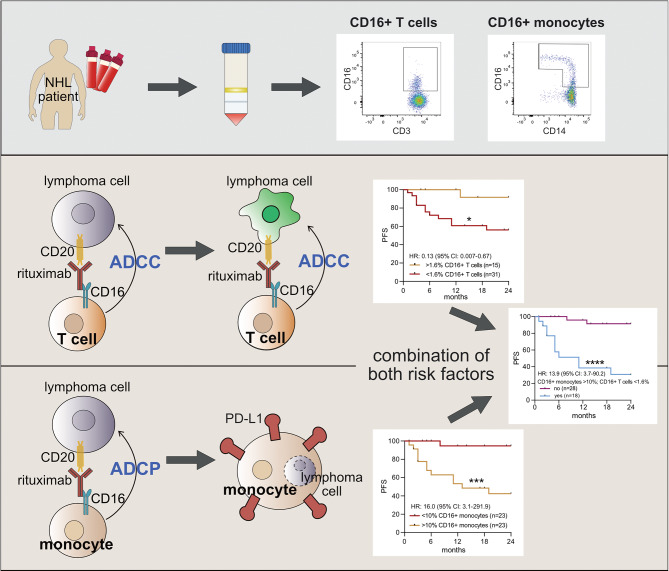
Assessing the prognosis of patients with aggressive non-Hodgkin B cell lymphoma mainly relies on a clinical risk score (IPI). Standard first-line therapies are based on a chemo-immunotherapy with rituximab, which mediates CD16-dependent antibody-dependent cellular cytotoxicity (ADCC). We phenotypically and functionally analyzed blood samples from 46 patients focusing on CD16+ NK cells, CD16+ T cells and CD16+ monocytes. Kaplan-Meier survival curves show a superior progression-free survival (PFS) for patients having more than 1.6% CD16+ T cells (p = 0.02; HR = 0.13 (0.007–0.67)) but an inferior PFS having more than 10.0% CD16+ monocytes (p = 0.0003; HR = 16.0 (3.1-291.9)) at diagnosis. Surprisingly, no correlation with NK cells was found. The increased risk of relapse in the presence of > 10.0% CD16+ monocytes is reversed by the simultaneous occurrence of > 1.6% CD16+ T cells. The unexpectedly strong protective function of CD16+ T cells could be explained by their high antibody-dependent cellular cytotoxicity as quantified by real-time killing assays and single-cell imaging. The combined analysis of CD16+ monocytes (> 10%) and CD16+ T cells (< 1.6%) provided a strong model with a Harrell’s C index of 0.80 and a very strong power of 0.996 even with our sample size of 46 patients. CD16 assessment in the initial blood analysis is thus a precise marker for early relapse prediction.
Graphical abstract
Supplementary Information
The online version contains supplementary material available at 10.1186/s12943-024-02123-7.
Keywords: aggressive B-NHL (non-Hodgkin B cell lymphoma), diffuse large B cell lymphoma (DLBCL), CD16+ T cell, CD16+ monocyte, antibody-dependent cellular cytotoxicity (ADCC), rituximab
Highlights
High CD16+ T cell counts have a positive correlation with PFS in aggressive NHL/DLBCL patients (p = 0.02; HR = 0.13, 0.01–0.7).
High CD16+ monocyte counts have a negative correlation with PFS in aggressive NHL/DLBCL patients (p = 0.0003; HR = 16.0, 3-292).
The combined assessment of CD16+ T cells and CD16+ monocytes accurately predicts PFS in aggressive NHL/DLBCL patients.
The strong protective function of CD16+ T cells could be explained by their high antibody-dependent cellular cytotoxicity.
Supplementary Information
The online version contains supplementary material available at 10.1186/s12943-024-02123-7.
Introduction
Diffuse large B cell lymphoma (DLBCL) is the most common type of aggressive lymphoma representing up to 40% of non-Hodgkin lymphomas (NHL). For more than 20 years, the standard first-line treatment for DLBCL patients has been immuno-chemotherapy with R-CHOP (rituximab, cyclophosphamide, doxorubicin, vincristine, and prednisolone) [1, 2] or similar approaches such as the recently approved pola-R-CHP (polatuzumab vedotin, rituximab, cyclophosphamide, doxorubicin, and prednisolone). This treatment regimen substitutes vincristine with the antibody-drug conjugate polatuzumab, which targets CD79b, and is recommended for patients with an International Prognostic Index (IPI) of 2–5 [3]. With these therapies, about 60–65% of high-risk patients can be cured, however refractory or relapsed (R/R) patients are characterized by a very poor outcome [4]. Therefore, reliable categorization of patients into low, intermediate, and high-risk categories is necessary to allow for allocation to different treatment regimens.
Risk stratification of patients with aggressive non-Hodgkin lymphoma (NHL), such as diffuse large B cell lymphoma (DLBCL), is mainly based on the clinical risk score IPI which was introduced 30 years ago [5] and includes the five adverse factors: age (> 60 years), Ann Arbor Stage (III/IV), more than one extranodal site, Eastern Cooperative Oncology Group (ECOG) working status (PS) of ≥ 2 and elevated level of lactate dehydrogenase. In addition, appropriate adequate immunophenotyping or gene expression profiling determines the cell of origin (CEO) (GCB vs. non-GCB). Although several modifications of the IPI have been proposed since then, the IPI remains the standard tool for risk stratification in newly diagnosed DLBCL patients and a better predictive model for personalized treatment has not been established yet [6].
Risk stratification must be simple, fast, and possible at the outset of diagnosis or prior to initiation of therapy to enable risk stratification for all patients without further loss of time. Here, we demonstrate that CD16 assessment of immune cell subsets in the initial blood analysis accurately predicts relapse within 24 months. Surprisingly, not CD16+ NK cell counts, but CD16+ T cell counts, a very small fraction of T cells, and CD16+ monocytes correlate with patient prognosis. CD16+ T cell counts have a positive correlation with PFS in aggressive B-NHL/DLBCL patients treated with R-CHOP, while CD16+ monocyte counts show a negative correlation. Finally, the combined assessment of the CD16+ T cell and CD16+ monocyte counts further improves the accuracy of relapse prediction. This information may help in the future to personalize therapy for specific patient groups.
Results
Patient characteristics
We conducted a prospective study analyzing peripheral blood samples from 46 patients with newly diagnosed non-Hodgkin lymphoma (NHL) at different stages of therapy. The majority of patients (85%) were diagnosed with diffuse large B cell lymphoma (DLBCL), 13% had other forms of high-grade NHL, and one patient had an aggressive form of follicular lymphoma that progressed to DLBCL. The median age of the patients at diagnosis was 67 years (range 22–91 years), and the median age of 25 sex- and age-matched healthy controls was 66 years (range 28–86 years). Patient details are shown in Table 1 (Supplementary Table 1). Of the patients, 59% had advanced disease (Ann-Arbor stage III-IV). According to the International Prognostic Index (IPI) score, 54% of patients were classified as low to intermediate risk (IPI 0–2) and 46% as intermediate to high risk (IPI 3–5).
The percentage of CD16+ T cells is a better predictor of progression-free survival than that of CD16+ NK cells
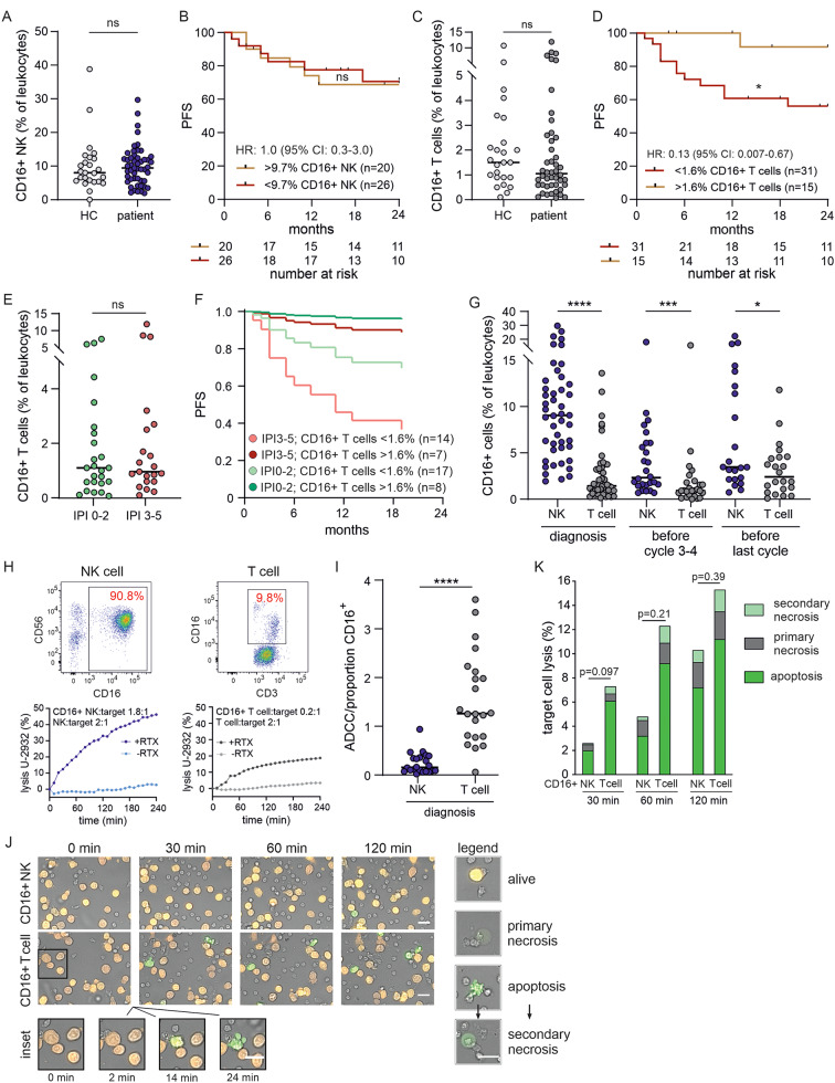
We performed flow cytometry analysis on purified peripheral blood mononuclear cells (PBMC) of newly diagnosed NHL patients to determine the percentage of CD16+ cells in peripheral blood leukocytes as all CD16+ cells can bind to rituximab and potentially affect the course of therapy. We found no difference in the percentage of CD16+ natural killer (NK) cells within the CD45 + leukocyte population between NHL patients and healthy controls (median 9.0% vs. 8.0%, p = 0.93; Fig. 1A). The analysis of progression-free survival (PFS) revealed no significant difference between patients with a high percentage (> 9.7%) and those with a low percentage (< 9.7%) of CD16+ NK cells (p = 0.86, HR = 1.0 (95% CI: 0.3-3.0); Fig. 1B). Next, we analyzed the percentage of CD16+ T cells in the CD45 + leukocyte population, which is only a small fraction of 1.5% in healthy patients (CD45 + CD3 + CD16+) and shows variable expression of CD4, CD8, and/or CD56 (Supplementary Fig. 1). Similar to the CD16+ NK cell population, we also found no difference in the percentage of CD16+ T cells between patients at diagnosis and healthy controls (median 1.1% vs. 1.5%, p = 0.27; Fig. 1C).
Fig. 1.
A high percentage of CD16+ T cells positively predicts patient outcome. A) Percentages of CD16+ NK in aggressive B-NHL patients (patient, n = 46) at diagnosis compared to healthy controls (HC, n = 25). B) Kaplan-Meier survival curve over 24 months. Progression-free survival (PFS) for high (> 9.7%) and low (< 9.7%) percentages of CD16+ NK cells at diagnosis. C) Percentages of CD16+ T cells in aggressive B-NHL patients at diagnosis (patient, n = 46) compared to healthy controls (HC, n = 25). D) Kaplan-Meier survival curve over 24 months. PFS for high (> 1.6%) and low (< 1.6%) percentages of CD16+ T cells at diagnosis. E) Percentages of CD16+ T cells at diagnosis according to patient’s IPI (IPI 0–2, n = 25; IPI 3–5, n = 21). F) Cox proportional hazard analysis of high (> 1.6%) and low (< 1.6%) percentages of CD16+ T cells at diagnosis in relation to IPI over 24 months. G) Percentages of CD16+ T cells and CD16+ NK cells at diagnosis and during therapy, (the number of patients (n) for each population (in the order given on the x-axes) n = 46, 46, 26, 26, 22, 22). H) Representative lysis capacity of NK and T cell populations (below) of one patient concerning their respective percentages of CD16+ cells (above). NK and T cells were isolated and lysis capacity was measured using a population-based real-time killing assay. The shown effector to target cell ratio (E: T) is 2:1. Percentages of CD16+ cells within the NK and T cell populations were quantified by flow cytometry. I) Quantification of antibody-dependent cytotoxicity (ADCC) in relation to the percentage of CD16+ NK (NK, n = 22) or CD16+ T cells (T cell, n = 22) within the whole NK or CD3 + T cell population. ADCC capacity corresponds to the difference between the lysis of target cells with rituximab (+ RTX) and without rituximab (-RTX). J) Representative images of rituximab-dependent ADCC by sorted CD16+ NK and CD16+ T cells at single cell level against TMD8 pCasper cells. Scale bars are 20 μm. K) Quantification of ADCC of sorted cells from four patients at different stages of therapy. HR hazard ratio; CI confidence interval; In Kaplan-Meier survival curves black marks represent censored patients; ns non-significant; * p < 0.05; *** p < 0.001; **** p < 0.0001
Compared to CD16+ NK cells, CD16+ T cells express significantly less CD16 as indicated by the median fluorescence intensity (MFI) (Supplementary Fig. 2). However, unexpectedly, we found that patients with a higher percentage of CD16+ T cells (> 1.6%) had a significantly better PFS as shown in the Kaplan-Meier survival curves (p = 0.02; HR = 0.13 (95% CI: 0.01–0.67); Fig. 1D). This prediction of PFS is not possible without the addition of CD16 (Supplementary Fig. 3). The additional analysis of the individual IPI score showed no difference in the percentage of CD16+ T cells in patients with low to intermediate risk (IPI 0–2) values compared to patients with intermediate to high risk (IPI 3–5) values (p = 0.51; Fig. 1E). In addition, Cox regression analysis confirmed that patients with a high percentage of CD16+ T cells (> 1.6%) have a clear PFS advantage, even when controlling for their individual IPI (Fig. 1F; high percentage of CD16+ T cells: HR = 0.12, p = 0.04; 95% CI: 0.02–0.93; high IPI: HR = 2.7, p = 0.08; 95% CI: 0.89–8.4). In patients with a low percentage of CD16+ T cells, those with high IPI scores showed worse PFS than those with low IPI scores (Fig. 1F). Thus, the elevated percentage of CD16+ T cells is an independent prognostic marker for improved PFS in patients with aggressive B-NHL/DLBCL.
CD16+ T cells are less sensitive to chemotherapy and exhibit a faster and more effective ADCC capacity than CD16+ NK cells
The remarkable observation that the percentage of CD16+ T cells has a greater impact on the PFS of patients than the percentage of CD16+ NK cells, raises the question how this could be explained. So far, it has been generally assumed that NK cells are the primary mediators of antibody-dependent cellular cytotoxicity (ADCC). This assumption is rationally based on the fact that the percentage of CD16+ NK cells is much higher than that of CD16+ T cells at the time of diagnosis (median 9.0% vs. 1.4%; Fig. 1G). In addition, we analyzed the percentage of CD16+ NK cells and CD16+ T cells not only at the time of diagnosis but also under therapy. These results show that the percentage of CD16+ NK cells decreases significantly until cycle 3–4 of R-CHOP therapy (p < 0.0001) with a large effect size of 1.12 (Cohen’s d) whereas the percentage of CD16+ T cells decreases to a lesser extent as demonstrated by the small effect size of 0.16 (Cohen’s d). Remarkably, the CD16+ T cell population recovers completely at the end of therapy unlike the CD16+ NK cells (Cohen’s d -0.16 vs. 0.44, respectively). As a consequence, the difference between the percentage of CD16+ NK cells and CD16+ T cells at the end of therapy was significantly smaller than at the beginning of therapy (median 9.0% CD16+ NK cells vs. median 1.4% CD16+ T cells; p < 0.0001 at diagnosis vs. median 3.5% CD16+ NK cells vs. median 2.4% CD16+ T cells, p = 0.05 at the end of therapy; Fig. 1G). Calculating the absolute counts of CD16+ NK and CD16+ T cells per µl of blood showed similar results (Supplementary Fig. 4). To gain functional insights into the potential mechanism by which CD16+ T cells are more relevant for the prognosis than CD16+ NK cells, we compared their respective efficiency regarding ADCC. For this purpose, we isolated NK and CD3 + T cells using magnetic beads and used them in an ADCC cytotoxicity assay. One representative example of an ADCC cytotoxicity assay of a patient’s entire NK and T cell population and the determination of the respective percentage of CD16+ cells within the total population analyzed by flow cytometry is shown in Fig. 1H. In the example of the NK population, 90% CD16+ NKs kill about 45% of the target cells after 4 h, whereas in the T cell population, only 10% CD16+ T cells kill about 20% of the target cells after 4 h. This means that the CD16+ T cells in this example are about 4 times more efficient. Based on the measured lysis capacity of the whole NK and T cell population and the percentage of CD16+ cells in the respective population, we calculated the ADCC capacity of the CD16+ cells for all patients, which were functionally analyzed. This calculation demonstrated that CD16+ T cells have an approximately 8-fold greater ADCC capacity than CD16+ NK cells (median 1.3 vs. 0.16; p < 0.0001; Fig. 1I). To further test the observation of increased cytotoxicity of CD16+ T cells, we sorted CD16+ T cells and CD16+ NK cells by flow cytometry for direct comparison at the single-cell level. As a target cell we used the DLBCL cell line TMD8 transduced with the GFP-RFP-FRET system (pCasper-GR). This assay allows single-cell analysis and distinguishes the mode of target cell death: apoptotic, primary necrotic or secondary necrotic [7]. The single-cell analysis of sorted CD16+ T cells and CD16+ NK cells demonstrated that the CD16+ T cells kill their target cells more efficiently by ADCC compared to the CD16+ NK cells as evidenced by green-fluorescent target cells (apoptotic cell death) and target cells which have lost their fluorescence (necrotic cell death) (Fig. 1J). Quantitative analysis reveals that this is primarily due to the faster kinetics of target cell lysis by the CD16+ T cells (Fig. 1K). We hypothesize that the positive correlation of a high percentage of CD16+ T cells on PFS is explained by a higher chemoresistance and an improved ADCC capacity of the CD16+ T cells compared to CD16+ NK cells.
Elevated levels of CD16+ monocytes are associated with reduced progression-free survival
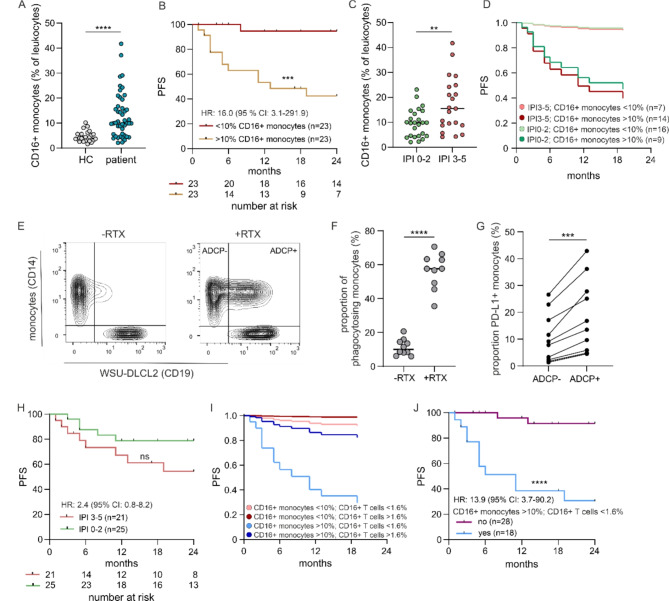
Unlike the percentage of CD16+ NK cells and CD16+ T cells which were similar between patients and healthy controls, the percentage of CD16+ monocytes (CD14 + + CD16+ (intermediate monocytes) or CD14 + CD16++ (nonclassical monocytes)) within the CD45+ leukocyte population was significantly higher in NHL patients at diagnosis compared to healthy controls (median 10.0% vs. 4%, p < 0.0001; Fig. 2A). Strikingly, patients with an increased percentage of CD16+ monocytes (> 10%) revealed a significantly worse PFS (p = 0.0003; HR = 16.0 (95% CI 3.1-291.9); Fig. 2B), which is not the case when analyzing the total monocyte population (Supplementary Fig. 3). Considering the patient’s individual IPI, we found that those patients classified as intermediate to high risk had significantly higher percentages of CD16+ monocytes compared to those classified as low to intermediate risk (median 15.7% vs. 9.9%, p = 0.003; Fig. 2C).
Fig. 2.
A high percentage of CD16+ monocytes negatively predicts patient outcome. A) Percentage of CD16+ monocytes (CD14 + + CD16+ (intermediate monocytes) or CD14 + CD16++ (nonclassical monocytes)) in aggressive B-NHL patients (patient, n = 46) at diagnosis compared to healthy controls (HC, n = 25). B) Kaplan-Meier survival curve over 24 months. Progression-free survival (PFS) for high (> 10%) and low (< 10%) percentages of CD16+ monocytes at diagnosis. C) Percentage of CD16+ monocytes at diagnosis according to patient’s IPI. (IPI 0–2, n = 25; IPI 3–5, n = 21). D) Cox proportional hazard analysis of high (> 10%) and low (< 10%) percentages of CD16+ monocytes at diagnosis in relation to IPI over 24 months. E) Representative staining of monocyte phagocytosis isolated from PBMC of a healthy donor. Monocytes were stained with anti-CD14 antibody and the target cell line WSU-DLCL2 with anti-CD19 antibody, co-cultured for 6 h without rituximab (-RTX) or with rituximab (+ RTX) and analyzed by flow cytometry. F) Percentage of monocytes that phagocytosed WSU-DLCL2 cells after co-culture of monocytes with WSU-DLCL2 cells for 6 h with (+ RTX) or without rituximab (-RTX); n = 10. G) Percentage of PD-L1+ monocytes after phagocytosis of WSU-DLCL2 cells (ADCP+) and monocytes without phagocytosing WSU-DLCL2 cells (ADCP-); n = 10. H) Kaplan-Meier survival curve over 24 months. PFS for patients with an IPI 0–2 or an IPI 3–5. I) Cox proportional hazard analysis of high (> 10%) and low (< 10%) percentages of CD16+ monocytes at diagnosis adjusted for high (> 1.6%) and low (< 1.6%) percentages of CD16+ T cells at diagnosis. CD16+ monocytes < 10%, CD16+ T cells < 1.6% (n = 13); CD16+ monocytes < 10%, CD16+ T cells > 1.6% (n = 10); CD16+ monocytes > 10%, CD16+ T cells < 1.6% (n = 18); CD16+ monocytes > 10%, CD16+ T cells > 1.6% (n = 5). J) Kaplan-Meier survival curve over 24 months. PFS for patients with a high percentage of CD16+ monocytes (> 10%) in combination with a low percentage of CD16+ T cells (< 1.6%) at diagnosis (light blue, yes) compared to patients without this combination (purple, no). In Kaplan-Meier survival curves, black marks represent censored patients. ns non-significant; ** p < 0.01; *** p < 0.001; **** p < 0.0001
To exclude the possibility that the IPI is not solely responsible for impaired PFS in patients with a high percentage of CD16+ monocytes, we conducted a Cox regression analysis adjusted for IPI (high percentage of CD16+ monocytes: HR = 14.5, p = 0.01; 95% CI: 1.8-116.5; high IPI: HR = 1.2, p = 0.7; 95% CI: 0.4–3.9; see also Fig. 2D). This analysis clearly shows that a higher percentage of CD16+ monocytes is correlated with a poorer PFS, with only a minor influence of the patient’s individual IPI. Patients with a low percentage of CD16+ monocytes (< 10%), showed a significantly better PFS even if their IPI classification was high and almost comparable to those with a low IPI (and a low percentage of CD16+ monocytes) (Fig. 2D). In conclusion, the percentage of CD16+ monocytes is a strong, independent prognostic marker for an impaired PFS in NHL patients. To gain functional insights why increased CD16+ monocyte counts predict poorer outcome, we performed a co-culture experiment with WSU-DLCL2 tumor cells to analyze rituximab-dependent phagocytosis (ADCP) of CD16+ monocytes. For this purpose, purified, fluorescently labeled monocytes from healthy donors were co-cultured for 6 h with fluorescently labeled tumor cells in the presence or absence of rituximab. Finally, we determined the percentage of tumor cells that were phagocytosed by the monocytes calculated on double-positive cells (Fig. 2E). As expected, we observed a significant increase in tumor cell phagocytosis by monocytes in the presence of rituximab (10.0% vs. 57.6%, p < 0.0001; Fig. 2F). However, we also observed that the percentage of PD-L1-positive monocytes significantly increased when monocytes had phagocytosed tumor cells (median 8.5% vs. 15.2% p = 0.0006; Fig. 2G). If rituximab administration would also correlate with upregulation of PD-L1 in CD16+ monocytes in vivo after rituximab-mediated tumor cell phagocytosis, NK and T cell activity would be inhibited. This in turn may explain the worse PFS of these patients due to the impairment of the key mediators of ADCC.
A high percentage of CD16+ T cells can overcome the reduced progression-free survival in patients with a high percentage of CD16+ monocytes
As the patient’s IPI is still the standard tool for risk stratification in newly diagnosed aggressive B-NHL/DLBCL patients, we analyzed PFS of patients dependent on their IPI. Kaplan-Meier survival curves show only a tendency but no significance for an impaired PFS in patients with higher IPI (Fig. 2H) which is most likely due to our small study size (n = 46). However, if we consider only high (4–5) and low (0–1) IPI, patients with high IPI (4–5) show a significantly impaired PFS (Supplementary Fig. 5, p = 0.01; HR 6.4; 95% CI 1.4–44.7). Importantly, this analysis does not provide any prognostic information for patients with intermediate IPI (2 or 3). Given the significant changes in patient’s PFS, depending on percentages of CD16+ T cells or CD16+ monocytes (Figs. 1D and 2B), we combined these two populations in a Cox regression analysis. This shows that only patients with a high percentage of CD16+ monocytes (> 10%) and a low percentage of CD16+ T cells (< 1.6%) have a significantly impaired PFS (Fig. 2I, J). A high percentage of CD16+ monocytes (> 10%) only slightly negatively influences the patient’s PFS if the percentage of CD16+ T cells is higher than 1.6%. The risk of an impaired patient’s PFS is low when both populations are low (CD16+ monocytes < 10% and CD16+ T cells < 1.6%). Patients with a high percentage of CD16+ T cells and a low percentage of CD16+ monocytes have almost no risk of a negative impact on PFS (Fig. 2I). It is noteworthy that only 5 out of 46 patients have a high percentage of CD16+ monocytes (> 10%) combined with a high percentage of CD16+ T cells (> 1.6%). This suggests that these subpopulations are not independent of each other. Noteworthy, even with only five patients in one group, the Kaplan-Meier survival analysis of the two groups (CD16+ monocytes > 10%; CD16+ T cells < 1.6% (n = 18) or CD16+ monocytes > 10%; CD16+ T cells > 1.6% (n = 5)) yielded a p-value of 0.07, which is close to the threshold of statistical significance. This further supports the conclusion that CD16+ T cells positively correlate with patient’s PFS. Combing the CD16+ T cell/CD16+ monocyte analysis with IPI did not enhance the prognostic value (Supplementary Fig. 6).
To emphasize the power of the combination of CD16+ T cells and CD16+ monocytes as a strong prognostic marker, we compared two groups in a Kaplan-Meier survival analysis. We combined in the one group patients with a high percentage of CD16+ monocytes (> 10%) and a low percentage of CD16+ T cells (< 1.6%) (n = 18, light blue). In the second group, we pooled all other possible combinations (n = 28, purple). The patients with a high percentage of CD16+ monocytes (> 10%) and a low percentage of CD16+ T cells (< 1.6%) have a median survival of only 11 months whereas 91% of the patients without this combination remained in remission until the end of our 24-month observation period (p = < 0.0001; Fig. 2J). The HR for patients with more than 10% CD16+ monocytes and less than 1.6% CD16+ T cells was 13.9 with a 95% CI of 3.7–90.2 (Fig. 2J). Furthermore, the Harrell’s C index was calculated to be 0.80 (95% CI: 0.71–0.88) indicating a good fit of our risk model.
In the present study, we offer new ways to accurately assess the risk of aggressive B-NHL/DLBCL patients at the time of diagnosis. In summary, even with the limited number of 46 patients, we have already achieved a power of 0.996 for the combined analysis of CD16+ monocytes and CD16+ T cells (α = 0.05).
The required analysis can be performed fast and relatively easy using standard hospital laboratory methods and thus offers the possibility to optimize personalized therapy at the time of diagnosis.
Discussion
This study has identified a novel prognostic marker for risk assessment in aggressive B-NHL/DLBCL patients. At the time of diagnosis, the combination of CD16+ monocytes and CD16+ T cells is a significant predictor of relapse probability, with a hazard ratio of 13.9 (95% CI: 3.7–90.2; p < 0.0001). The combined analysis of CD16+ monocytes (> 10%) and CD16+ T cells (< 1.6%) resulted in a very strong power of 0.996 even with our relatively small sample size of 46 patients. However, we see two limitations: (1) The study was performed using peripheral blood mononuclear cells (PBMCs) purified from blood samples. Future studies should be conducted using whole blood analyses. This approach would facilitate standardization while minimizing laboratory time and costs at the time of diagnosis. (2) Validation of the results in a larger patient cohort is not yet available.
The required numbers can be determined as part of the standard blood collection from patients at the time of diagnosis, with the simple additional step of flow cytometric staining, which includes the antibodies CD45, CD14, CD3, and CD16. While the combined analysis of CD16+ monocytes and CD16+ T cells is indicative of the relapse probability, the percentage of CD16+ T cells alone (> 1.6% CD16+ T cells) is a protective predictor for the PFS of patients (over 24 month).
A high amount of monocytes has long been discussed as a marker of poor patient outcome for hematological and non-hematological malignancies [8, 9]. Similar results were found in further studies [10–12]. In these studies, the analysis of the patients’ blood did not distinguish whether rituximab was added to the therapy or not, and the number of CD16+ monocytes was not determined separately. Reviewing 1,057 DLBCL patients revealed that a high lymphocyte/monocyte ratio was highly predictive of better outcome and event-free survival only in those patients who also received rituximab compared to chemotherapy alone [13]. Subsequently, it was demonstrated that CD16+ monocyte subtypes can also predict the prognosis of DLBCL patients. In a study with a comparable number of patients, Han et al. found a significantly increased number of CD16+ monocytes in DLBCL patients. Le Gallou et al. analyzed monocyte subtypes in more detail and also found an elevated proportion of nonclassical monocytes (CD14+/CD16++) in the peripheral blood to be negatively associated with an adverse prognosis of DLBCL patients [14].
However, to the best of our knowledge, no studies have yet been published on the specific function of CD16+ T cells in aggressive B-NHL/DLBCL and there is currently no evidence of any association between the presence of CD16+ T cells and the progression-free survival of aggressive B-NHL/DLBCL patients. However, it was shown that high frequencies of CD16-expressing γδ T cell infiltration in tumor tissue positively correlates with clinical outcome [15].
Our findings demonstrate a protective role for the CD16+ T cell population in patients with aggressive B-NHL. Specifically, we found that patients with more than 1.6% CD16+ T cells (as a percentage of CD45 + leukocytes) at diagnosis had not only a significantly better PFS (Fig. 1D) but were also able to overcome impairment of PFS for patients with a high proportion of CD16+ monocytes (Fig. 2I, light and dark blue curves).
The combined analysis of both subpopulations, CD16+ T cells and CD16+ monocytes, has the potential to offer a more personalized medicine for patients with newly diagnosed aggressive B-NHL/DLBCL. The results of this study provide a rationale for investigating whether patients diagnosed with aggressive B-NHL/DLBCL could receive either reduced or more intensive therapy, depending on the results of flow cytometry at the initial blood analysis simply by adding the anti-CD16 antibody to the respective standard panel. This addition of anti-CD16 antibodies allows a precise statement as to whether patients have a relapse (CD16+ monocytes > 10%, CD16 T cells < 1.6%) or a dramatically reduced risk of relapse (CD16+ monocytes < 10%, CD16+ T cells > 1.6%).
Electronic supplementary material
Below is the link to the electronic supplementary material.
Acknowledgements
We thank Kathleen Seelert for excellent technical help. We are grateful to Prof. Dr. Stephan Stilgenbauer (CCCU, Ulm University, Germany) for his continuous support. We are grateful to all human blood donors.
Author contributions
SZ, NK, JJ, CH, GS (Schäfer) performed experiments. JJW performed statistical analysis. SZ, LK, GS did statistical analysis. JV, LK, GS established single cell analysis. HE, PO provided samples from healthy donors. CS, FN organized samples from B-NHL /DLBCL patients. DY supported with cell sorting. AM, PW, EU generated and conceptualized pCasper target cell lines. OC, MB, LT selected and evaluated clinical samples. SZ, MH, MB, LT, ECS conceptualized and implemented the project. SZ planned experiments. SZ, NK, ECS analyzed the data. SZ designed the figures, SZ and ECS wrote the manuscript. All authors were involved in the critical review of the manuscript. All authors read and approved the manuscript.
Funding
This work was supported by the Deutsche Forschungsgemeinschaft (DFG, the collaborative research centers SFB 1027 (project A11 to MH), SFB 894 (project A1 to MH), DFG-State Major Instrumentation, GZ: INST256/429-1 FUGB (Molecular Devices, High content Screening System) and GZ: INST 256/423-1 FUGG (BD Biosciences, FACSVerse), and by the Bundesministerium für Bildung und Forschung (BMBF, grant 031LO133 to MH)). EU received funding by the Stiftung Deutsche Krebshilfe (German Cancer Aid; #70114180 and #70115200); EU and AM were supported by DFG (4D-CARLY - UL 316/9 − 1 and SFB/CRC1292, Project-ID 318346496).
Data availability
No datasets were generated or analysed during the current study.
Declarations
Ethical approval
The study on aggressive B-NHL/DLBCL patients (Internal Medicine I, Saarland University), healthy volunteers (Institute for Clinical and Experimental Surgery, Saarland University) and the research with human PBMC derived from leukocyte reduction system (LRS) chambers have been approved by the local ethic committee (33/18 Prof. Dr. Grundmann, 84/15 Prof. Dr. Rettig-Stürmer). Written consent for the use of their blood in research was obtained from patients (Internal Medicine I, Saarland University) and healthy volunteers.
Competing interests
The authors declare no competing interests.
Footnotes
Publisher’s note
Springer Nature remains neutral with regard to jurisdictional claims in published maps and institutional affiliations.
References
- 1.Coiffier B, Lepage E, Briere J, Herbrecht R, Tilly H, Bouabdallah R, Morel P, Van Den Neste E, Salles G, Gaulard P, et al. CHOP chemotherapy plus rituximab compared with CHOP alone in elderly patients with diffuse large-B-cell lymphoma. N Engl J Med. 2002;346:235–42. [DOI] [PubMed] [Google Scholar]
- 2.Pfreundschuh M, Trumper L, Osterborg A, Pettengell R, Trneny M, Imrie K, Ma D, Gill D, Walewski J, Zinzani PL, et al. CHOP-like chemotherapy plus rituximab versus CHOP-like chemotherapy alone in young patients with good-prognosis diffuse large-B-cell lymphoma: a randomised controlled trial by the MabThera International Trial (MInT) group. Lancet Oncol. 2006;7:379–91. [DOI] [PubMed] [Google Scholar]
- 3.Tilly H, Morschhauser F, Sehn LH, Friedberg JW, Trneny M, Sharman JP, Herbaux C, Burke JM, Matasar M, Rai S, et al. Polatuzumab Vedotin in previously untreated diffuse large B-Cell lymphoma. N Engl J Med. 2022;386:351–63. [DOI] [PubMed] [Google Scholar]
- 4.Sehn LH, Salles G. Diffuse large B-Cell lymphoma. N Engl J Med. 2021;384:842–58. [DOI] [PMC free article] [PubMed] [Google Scholar]
- 5.International Non-Hodgkin’s Lymphoma Prognostic Factors P. A predictive model for aggressive non-hodgkin’s lymphoma. N Engl J Med. 1993;329:987–94. [DOI] [PubMed] [Google Scholar]
- 6.Jelicic J, Larsen TS, Maksimovic M, Trajkovic G. Available prognostic models for risk stratification of diffuse large B cell lymphoma patients: a systematic review. Crit Rev Oncol Hematol. 2019;133:1–16. [DOI] [PubMed] [Google Scholar]
- 7.Backes CS, Friedmann KS, Mang S, Knorck A, Hoth M, Kummerow C. Natural killer cells induce distinct modes of cancer cell death: discrimination, quantification, and modulation of apoptosis, necrosis, and mixed forms. J Biol Chem. 2018;293:16348–63. [DOI] [PMC free article] [PubMed] [Google Scholar]
- 8.Gu L, Li H, Chen L, Ma X, Li X, Gao Y, Zhang Y, Xie Y, Zhang X. Prognostic role of lymphocyte to monocyte ratio for patients with cancer: evidence from a systematic review and meta-analysis. Oncotarget. 2016;7:31926–42. [DOI] [PMC free article] [PubMed] [Google Scholar]
- 9.Wen S, Chen N, Peng J, Ling W, Fang Q, Yin SF, He X, Qiu M, Hu Y. Peripheral monocyte counts predict the clinical outcome for patients with colorectal cancer: a systematic review and meta-analysis. Eur J Gastroenterol Hepatol. 2019;31:1313–21. [DOI] [PubMed] [Google Scholar]
- 10.Li YL, Gu KS, Pan YY, Jiao Y, Zhai ZM. Peripheral blood lymphocyte/monocyte ratio at the time of first relapse predicts outcome for patients with relapsed or primary refractory diffuse large B-cell lymphoma. BMC Cancer. 2014;14:341. [DOI] [PMC free article] [PubMed] [Google Scholar]
- 11.Maurer MJ, Jais JP, Ghesquieres H, Witzig TE, Hong F, Haioun C, Thompson CA, Thieblemont C, Micallef IN, Porrata LF, et al. Personalized risk prediction for event-free survival at 24 months in patients with diffuse large B-cell lymphoma. Am J Hematol. 2016;91:179–84. [DOI] [PMC free article] [PubMed] [Google Scholar]
- 12.Tadmor T, Bari A, Sacchi S, Marcheselli L, Liardo EV, Avivi I, Benyamini N, Attias D, Pozzi S, Cox MC, et al. Monocyte count at diagnosis is a prognostic parameter in diffuse large B-cell lymphoma: results from a large multicenter study involving 1191 patients in the pre- and post-rituximab era. Haematologica. 2014;99:125–30. [DOI] [PMC free article] [PubMed] [Google Scholar]
- 13.Rambaldi A, Boschini C, Gritti G, Delaini F, Oldani E, Rossi A, Barbui AM, Caracciolo D, Ladetto M, Gueli A, et al. The lymphocyte to monocyte ratio improves the IPI-risk definition of diffuse large B-cell lymphoma when Rituximab is added to chemotherapy. Am J Hematol. 2013;88:1062–7. [DOI] [PubMed] [Google Scholar]
- 14.Han X, Ruan J, Zhang W, Zhou D, Xu D, Pei Q, Ouyang M, Zuo M. Prognostic implication of leucocyte subpopulations in diffuse large B-cell lymphoma. Oncotarget 2017;8:47790–47800. 10.18632/oncotarget.17830 [DOI] [PMC free article] [PubMed]
- 15.Le Gallou S, Lhomme F, Irish JM, Mingam A, Pangault C, Monvoisin C, Ferrant J, Azzaoui I, Rossille D, Bouabdallah K, et al. Nonclassical monocytes are Prone to migrate into tumor in diffuse large B-Cell lymphoma. Front Immunol. 2021;12:755623. [DOI] [PMC free article] [PubMed] [Google Scholar]
- 16.Saura-Esteller J, de Jong M, King LA, Ensing E, Winograd B, de Gruijl TD, Parren P, van der Vliet HJ. Gamma Delta T-Cell Based Cancer Immunotherapy: Past-Present-Future. Front Immunol. 2022;13:915837. [DOI] [PMC free article] [PubMed] [Google Scholar]
Associated Data
This section collects any data citations, data availability statements, or supplementary materials included in this article.
Supplementary Materials
Data Availability Statement
No datasets were generated or analysed during the current study.


