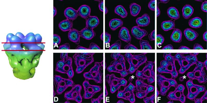
FIG. 7.
Antibody-induced structural changes in the virion. Sections (14-Å thick) through the ς3 protein layer (top row) at a radius of ∼407 Å and through the μ1 protein layer (bottom row) at a radius of ∼385 Å are displayed for each reconstruction. Red lines indicate the locations of these sections on a side view of a conical cutaway of the hexameric arrangement of ς3 and μ1 in native virions. In native virions (A), ς3 subunits appear to be in contact with each other, whereas in the IgG-bound virions (B) and Fab-bound virions (C), the ς3 subunits are more distinct. In native virions, an unobstructed channel is observed traversing the μ1 layer (D). However, in IgG-bound virions (E) and Fab-bound virions (F), a small density is present at the center of the hexameric arrangement of μ1 proteins (∗).

