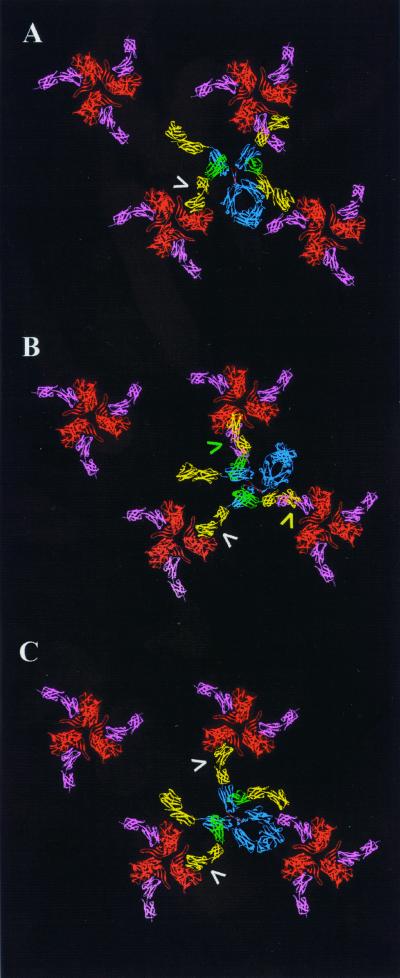
FIG. 3.
Models of CD4-IgG2 in complex with arrays of gp120 trimers. (A) Docking of CD4-IgG2 with trimers oriented such that the CD4 binding sites project toward neighboring trimers. (B and C) gp120 trimers are rotated 20° and 45° clockwise with respect to those in panel A. Arrows indicate CD4 arms that closely approach (yellow and green) or fully engage (white) a CD4 binding site of gp120. Structures are color coded as described for Fig. 2.

