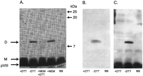
FIG. 2.
SDS-PAGE analysis of the f88-4 wild-type phage (f88) and recombinant B2.1 phage (all others). Phage were left untreated or treated with DTT, NEM, or NEM followed by DTT and then analyzed by SDS-PAGE. Monomeric (M) and dimeric (D) recombinant pVIII proteins are shown. Proteins in similar gels were either silver stained or transferred to a membrane and subjected to Western blotting with anti-phage Ab or IgG1 b12. Panels: A, silver-stained gel; B, Western blot using IgG1 b12 to show the reactive dimer; C, Western blot using rabbit anti-phage Ab to show the wild-type and recombinant pVIII proteins.

