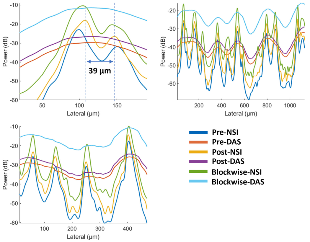
Fig. 15.

Microvessel cross sectional profiles of PD microvessel images of contrast enhanced rat brain. Top left: first cross section marked with blue solid line in Fig.14; Top right: second cross section marked with orange solid line in Fig.14; Bottom left: third cross section marked with yellow solid line in Fig.14.
