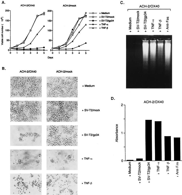
FIG. 2.
Detection of apoptosis of the ACH-2/OX40 cells stimulated with gp34. (A) ACH-2/OX40#10 cells and ACH-2/mock#7 cells were cultured for 5 days with medium alone, PFA-fixed SV-T2/mock cells, PFA-fixed SV-T2/gp34 cells, TNF-α, or TNF-β and then examined for viable cell number under a microscope. The results are expressed as mean numbers of living cells obtained in triplicate. (B) Morphological alteration of ACH-2OX40#10 cells and ACH-2/mock#7 cells 3 days after the introduction of various stimuli. Original magnification, ×200. (C) Nuclear DNA fragmentation of ACH-2/OX40#10 cells (106) by apoptosis was analyzed by 2% agarose gel electrophoresis 3 days after the stimuli. Anti-Fas MAb (CH-11) was used as positive control. (D) The apoptotic DNAs of ACH-2/OX40#10 cells (103/well) were measured by using an ELISA kit 3 days after various stimuli. The results are a mean number of absorbance values obtained in triplicate. Representative results are shown from two independent experiments.

