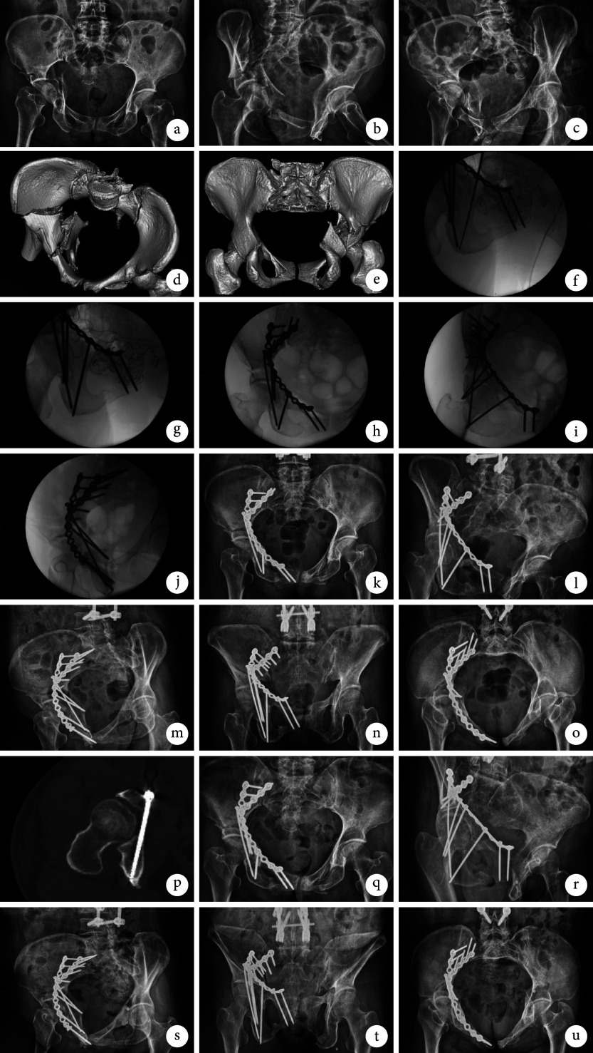
图 2.
A 56-year-old female patient with right acetabular fracture (Letournel-Judet classifcation: T-shaped fracture)
患者,女,56岁,右侧髋臼骨折(Letournel-Judet分型为 T型骨折)
a~c. 术前骨盆前后位、右侧闭孔斜位及右侧髂骨斜位X线片;d、e. 术前骨盆CT三维重建;f、g. 术中IAS植入过程; h~j. 术中骨盆前后位、右侧闭孔斜位、右侧髂骨斜位透视示髋臼骨折复位满意,IAS位置良好;k~o. 术后7 d骨盆前后位、右侧闭孔斜位、右侧髂骨斜位、出口位及入口位X线片示髋臼骨折复位满意,IAS位置良好;p. 术后8 d CT示IAS全程位于通道内;q~u. 术后3个月骨盆前后位、右侧闭孔斜位、右侧髂骨斜位、出口位及入口位X线片示骨折愈合
a-c. Preoperative X-ray films of pelvic anteroposterior view, right obturator oblique view, and right iliac oblique view; d, e. Preoperative three-dimensional reconstruction of pelvic CT scan; f, g. Intraoperative implantation of IAS; h-j. Intraoperative fluoroscopy of pelvic anteroposterior view, right obturator oblique view, and right iliac oblique view showed that the reduction of acetabular fracture was satisfactory and the position of IAS was good; k-o. At 7 days after surgery, X-ray films of pelvic anteroposterior view, right obturator oblique view, right iliac oblique view, outlet view, and inlet view showed that the reduction of acetabular fracture was satisfactory, and the position of IAS was good; p. At 8 days after surgery, CT scan showed that the IAS trajectory was located in the channel; q-u. At 3 months after surgery, X-ray films of pelvic anteroposterior view, right obturator oblique view, right iliac oblique view, outlet view, and inlet view showed that the fracture healed

