. 2024 Sep 28;12(9):e008568. doi: 10.1136/jitc-2023-008568

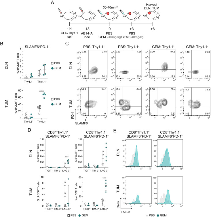
Figure 3. Chemotherapy induced increase in SLAMF6+PD-1+ CD8+ TPEX is not restricted to tumor antigen-specific CD8+ T cells. (A) Experiment timeline. BALB/c recipients received CL4xThy1.1 splenocytes 1-day prior to AB1-HA tumor inoculation. Mice were treated with two doses of either PBS or GEM when tumors reached 30–40 mm2 in size. Tumors (TUM) and tumor draining lymph nodes (DLN) were harvested for flow cytometry 3 days after the second dose of chemotherapy (+6). (B) Dot plots showing the percentage of Thy1.1+ or Thy1.1 SLAMF6+PD-1+ TPEX out of CD8+ T cells in PBS and GEM treated DLNs (top) and TUM (bottom). (C) Representative flow cytometry plots of SLAMF6 and PD-1 expression on CD8+Thy1.1+ and CD8+Thy1.1 T cells in DLNs (top) and TUM (bottom). (D) Dot plots presenting the frequency of TIGIT, TIM-3 and LAG-3 expression on CD8+Thy1.1+ (left) and CD8+Thy1.1 (right) T cells in DLNs (top) and TUM (bottom). (E) Representative histograms of LAG-3 expression on CD8+Thy1.1+ (left) and CD8+Thy1.1 (right) TPEX in DLNs and TUM. Data represented as mean±SD. Two-way analysis of variance with Tukey’s multiple comparisons test was used to compare between treatment groups and cell types. Sample sizes n=4 per treatment group. *p<0.05, **p<0.01, ****p≤0.0001. GEM, gemcitabine; LAG-3, lymphocyte activating gene-3; PBS, phosphate-buffered saline; PD-1, programmed cell death protein 1; SALMF6, SLAM family member 6; TIGIT, T-cell immunoreceptor with immunoglobulin and ITIM domain; TIM-3, T-cell immunoglobulin and mucin-domain containing-3; TPEX, progenitor exhausted CD8+ T cells.

Figure 3