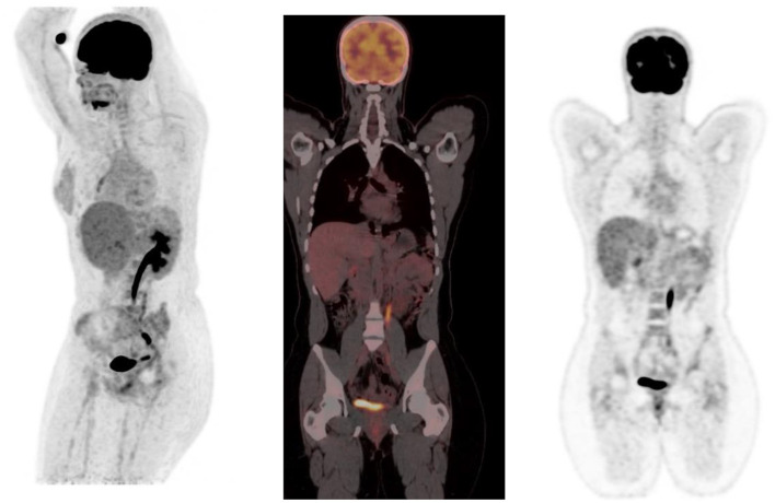
Figure 14.
MIP and coronal FDG PET/CT and PET images showing physiological activity in the left renal collecting system, left ureter and urinary bladder. The MIP image is helpful in differentiating tracer excretion in the ureter from nodal pathology. Identifying the uptake to the ureter on CT also helps in differentiating from nodal pathology.

