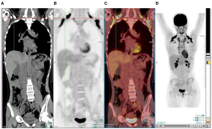
Figure 16.
15 year old male with stage 3 Hodgkin's lymphoma referred for follow up FDG PET post chemotherapy. Coronal PET (B), and PET/CT (C) showing bilateral symmetrical uptake in the neck and paravertebral region of the cervical and thoracic spine. Crosshairs localized to fat density on CT (A). FDG uptake in brown adipose tissue is commonly seen in children and adolescents. Many pediatric patients have mild brown fat uptake in the neck or supraclavicular regions. However, intense uptake may include pericardiac and perirenal brown adipose tissue. 18F-FDG uptake in activated brown fat may obscure sites of pathologic FDG uptake and decrease confidence in the interpretation of the study.

