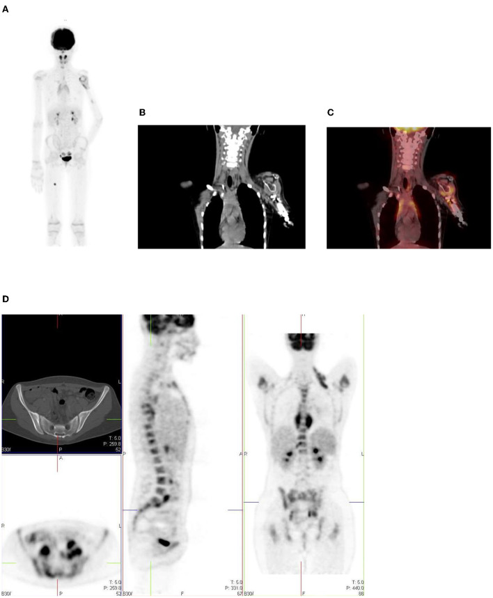
Figure 17.
(A–C) 9 year old female with high grade osteosarcoma. Coronal PET image (A) shows normal pediatric red marrow distribution. There is homogenous FDG uptake in the proximal humeri, proximal and distal femurs, and proximal tibias reflecting normal FDG uptake in red bone marrow. This uptake is usually minimal or absent in adults due to the conversion of red marrow to yellow marrow, which is less metabolically active. Pathology in the left shoulder with increased FDG uptake in the left humeral head and proximal humerus is compatible with orthopedic hardware (B). CT image (B) showing the prosthesis and fused PET/CT image (C) showing the peri-prosthetic FDG uptake. Note however, active disease cannot entirely be excluded. Due to pain in the left arm, the patient was unable to extend and rotate the left hand for correct imaging position. (D) 16 year old female with newly diagnosed Hodgkin's lymphoma. Staging PET shows increased uptake in the marrow, greater than the liver with areas of inhomogeneous and focal uptake, indicating bone marrow infiltration. The CT did not show anatomical lesions (Top left). Note the left neck (site of biopsy confirmed disease) and intrathoracic nodal disease.

