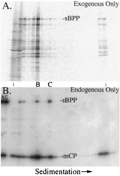
FIG. 4.
SCMV BPP binds to capsids in lysates of SCMV-infected cells. The nuclear fractions of SCMV-infected cells were incubated with 35S-sBPP, the mixtures were separated by rate-velocity centrifugation, and gradient fractions were subjected to SDS-PAGE followed by electrotransfer to Immobilon membranes. Shown are phosphorimages prepared from the membrane before (A) and after (B) it was probed with a mixture of anti-sBPP and anti-mCP. An acetate sheet and a sheet of XAR film were placed between the membrane and imaging plate to block the 35S signal for panel B. B- and C-capsids (denoted between the panels) were determined to be in fractions 6 and 9, respectively, by the pattern of CBB-stained proteins in a separate gel (not shown) and by the peak intensities of mCP following immunoassay (B). NP-40 nuclear (N) and cytoplasmic (C) fractions of nonradiolabeled SCMV-infected cells were included in the leftmost lanes as position markers for the protein bands of interest. The vertical lines between the panels indicate the first and last fractions of the gradient.

