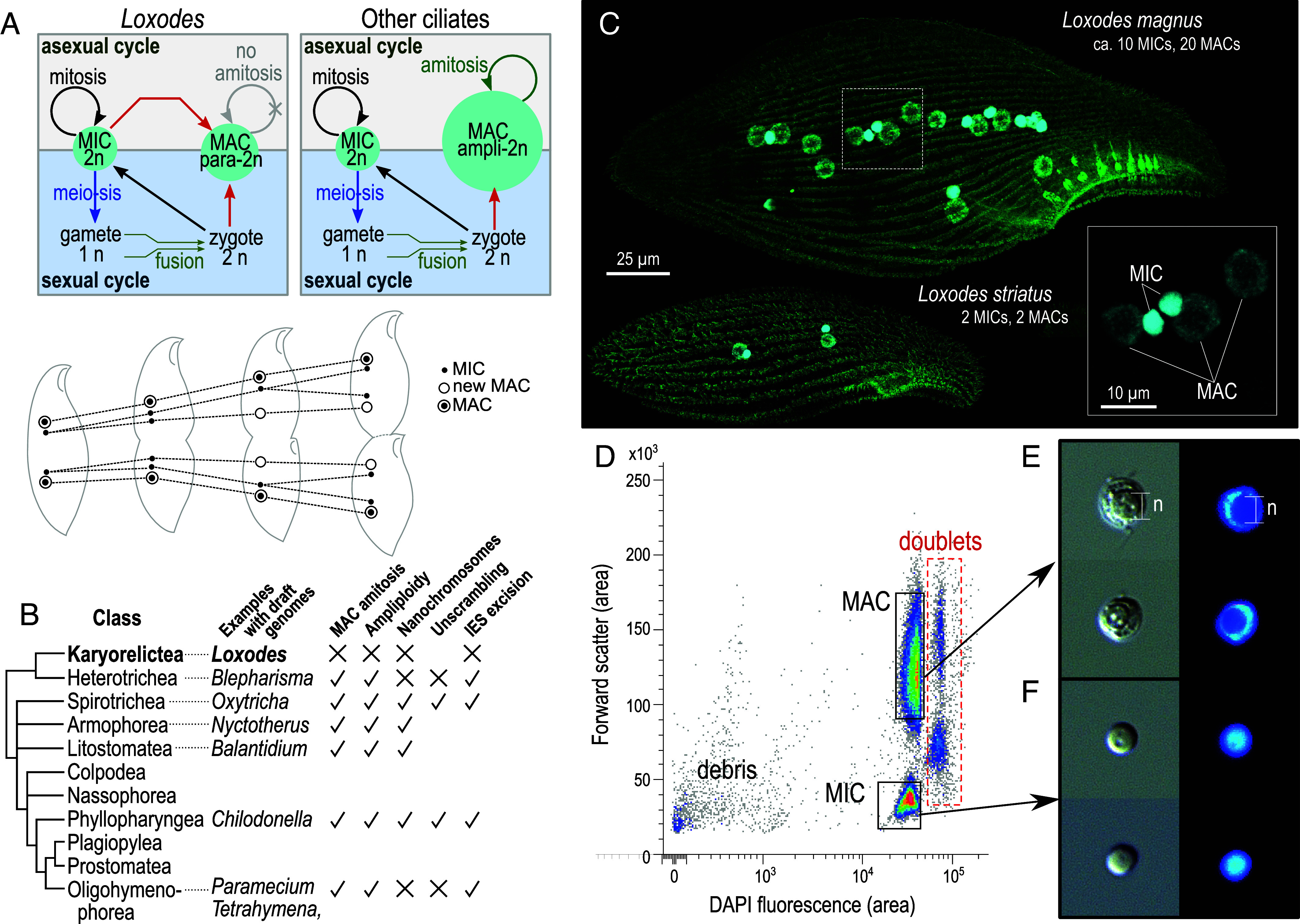
Fig. 1.

Loxodes nuclei purification. (A) Simplified diagram of nucleus development in typical ciliates vs. Loxodes and other karyorelicts (above); nuclei in L. striatus during asexual division, after (35) (below). (B) Diagrammatic tree of ciliate classes [after (37), branch lengths arbitrary] and genome architecture characteristics with evidence from draft genomes. (C) Confocal scanning fluorescence micrographs of Loxodes cells (maximum-intensity projections): green, alpha-tubulin secondary immunofluorescence; cyan, DAPI staining of nuclei; Inset, detail of L. magnus nuclei. (D) Representative flow cytometric scatterplot of forward scatter vs. DAPI fluorescence for L. magnus cell lysate (39,312 events depicted), with gates for MAC and MIC defined for flow sorting. Median integrated DAPI fluorescence for MACs was 116% that of MICs. (E and F) MAC and MIC respectively after sorting, imaged with differential interference contrast (Left) and DAPI fluorescence (Right); each subpanel width 10 µm. The spherical nucleolus (“n”) is less densely stained (panel E).
