Figure 2.
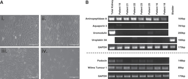
Urine-derived cell characterization study results. (A) Representative images of different cell morphologies observed in urine-derived cells. Variation in cell morphology was observed, with multiple types sometime present within 1 sample. Most commonly encountered proliferating types are (i–iii) with (iv) never reaching confluency. Cells with rice-grain morphology (iii) exhibited the best proliferative capacity, eventually becoming elongated (ii) and uniform with repeat passaging. Scale bar=50 µm. (B) RT-PCR result summary. Tubulopathy patient-derived urinary cells were characterized using RT-PCR to detect transcripts of ENPEP (aminopeptidase A), AQP3 (aquaporin 3), UMOD (uromodulin), UPK3A (uroplakin 3A), NPHS2 (podocin), and WT1 (Wilms tumor 1). ENPEP and WT1 transcripts were detected in all patient samples.

