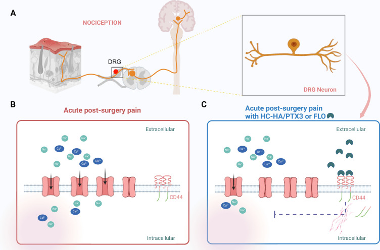
Figure 1. Targeting pain signaling at its source.
(A) Painful stimuli are detected through a process called nociception. Neurons (orange) located in the dorsal root ganglia (DRG) relay pain signals from a peripheral wound (such as a skin injury; left) to the central nervous system (right), which results in pain being experienced. (B) After surgery, sodium ions (Na+; green) and calcium ions (Ca2+; blue) flow into DRG nociceptive neurons through ion channels (pink) that are embedded in the cell wall of the neurons. This contributes to acute post-surgical pain. (C) HC-HA/PTX3 or FLO (dark green shapes) can bind to CD44 receptors (red lines) on the surface of the DRG neurons. This binding leads to a rearrangement of the cytoskeleton within the cell, which pushes the receptors into the neuron, causing some of the ion channels to close. The resulting reduction in the influx of sodium and calcium ions leads to a decrease in pain signaling. This figure was created using BioRender.com.

