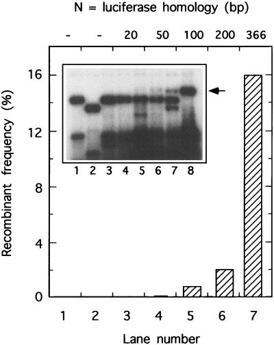
FIG. 2.
Southern blot analysis of recombinant molecules recovered from vaccinia virus-infected cells. DNA was recovered from cells transfected with the luciferase-encoding plasmids indicated in the legend to Fig. 1, digested with SpeI, XhoI, and DpnI (to degrade unreplicated input DNA), and size fractionated. After transfer to a nylon membrane, plasmid sequences were detected with a 32P-labeled luciferase probe and the yield of 1.7-kbp recombinant molecules (indicated by the arrow) was determined by densitometry. Cells were also transfected with a plasmid carrying an intact luciferase gene (lane 8), and the recombinant frequencies were calculated relative to the intensity of the 1.7-kbp full-length luciferase-encoding fragment seen in that lane. No recombinant DNA was detected in cells transfected with pRP406Δ or pRP403Δ alone (lanes 1 and 2, respectively). Note that different vectors were used to construct pXYT403Δ20 to pXYT403Δ200 (lanes 3 through 6) versus those for pRP403Δ (lanes 2 and 7). Recombinant frequencies can still be calculated because the luciferase-encoding recombinant fragments are identical, but differences in flanking vector sequences produce other changes in the restriction patterns.

