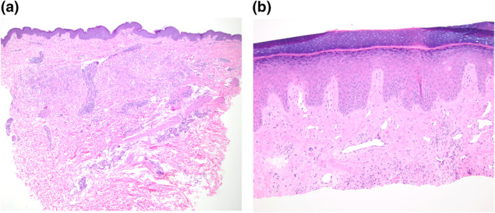
FIGURE 2.

(a) Histopathology of a left flank skin lesion showing an interstitial and palisaded histiocytic infiltrate surrounding degenerated collagen in the dermis, consistent with granuloma annulare (Case 1). (b) Shave biopsy from the dorsal hand showed the upper portion of an interstitial histiocytic infiltrate compatible with interstitial GA (Case 2). Magnifications: 4X(a), 10X(b).
