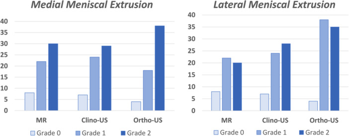
Figure 2.

Semi‐quantitative grading of medial and lateral meniscal extrusion based on the three different imaging evaluations: magnetic resonance (MR), clinostatic ultrasound (clino‐US) and orthostatic ultrasound (ortho‐US).

Semi‐quantitative grading of medial and lateral meniscal extrusion based on the three different imaging evaluations: magnetic resonance (MR), clinostatic ultrasound (clino‐US) and orthostatic ultrasound (ortho‐US).