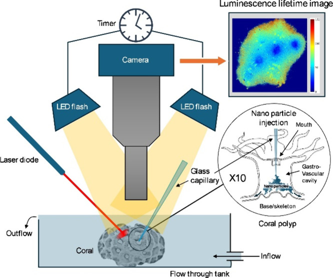
Figure 1.

Schematic drawing of the setup used for injecting O2 sensor nanoparticles into the gastrovascular cavity of coral polyps and subsequent mapping of the internal O2 concentration with a luminescence lifetime system. The inset shows a coral polyp, where a glass microcapillary is introduced through the coral mouth into the gastrovascular cavity for injection of sensor nanoparticles that can then distribute to neighboring polyps via channels in the connective tissue (see also SVideo 1). A laser pointer was used to locally stimulate photosynthetic O2 production.
