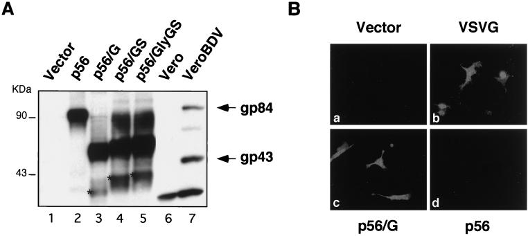
FIG. 2.
Expression of wild-type and chimeric GPs. (A) Detection of wild-type BDV p56 and chimeric GPs in cell lysates. BHK-21 cells were transfected with the indicated plasmids. Forty-eight hours after transfection cells were lysed in sample buffer. Protein lysates were separated by SDS–8 to 16% PAGE and analyzed by Western blotting using an anti-BDV p56 rabbit serum. Vero cells mock infected (lane 6) or persistently infected with BDV (lane 7) were used as negative and positive controls, respectively. Arrows, migrations of the full-length (gp84) and processed (gp43) BDV GPs; asterisks, positions of the nonglycosylated precursors. (B) Intracellular staining of cells transiently expressing VSV G and BDV-VSV chimeric GPs. After 48 h, BHK-21 cells transfected with the indicated plasmid were fixed and permeabilized with methanol-acetone. Cells were stained with a rabbit serum that specifically recognizes the CT of the VSV GP. The results with p56/G (c) are shown as a representative example. Similar results were obtained with p56/GS and p56/GlyGS. The antibody anti-VSV CT did not cross-react with BDV p56 (d).

