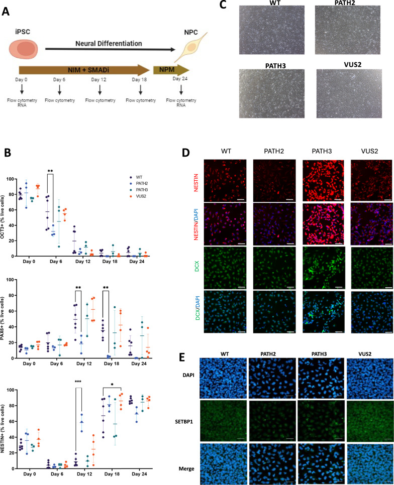
Fig. 2.
Differentiation of SETBP1 iPSC clones into neural progenitor cells. A Neural differentiation schematic showing differentiation of iPSCs into neural progenitor cells (NPCs) using neural induction media (NIM) and SMADi followed by neural progenitor media (NPM). Cells were harvested at selected timepoints across differentiation for pluripotency and neural marker analysis using flow cytometry and RNAseq. Image generated using BioRender. B Pluripotency and neural marker expression across neural differentiation. Graph plots indicate OCT3, PAX6 and NESTIN expression was assessed by flow cytometry in differentiating iPSC clones at 6-day intervals. Data presented as mean ± s.d.*p < 0.05; **p < 0.01; ***p < 0.001. WT: wild-type. C Morphology of NPCs derived from SETBP1 gene-edited iPSC clones. Representative images of NPCs harbouring PATH2, PATH3, VUS2 SETBP1 variants and WT NPCS at 4X objective magnification. D Representative images of neural marker, NESTIN and DCX, immunostaining in NPCs harbouring SETBP1 variants. Scale bar 50 µm. E Representative images of SETBP1 staining (green) relative to nuclear staining (blue) in iPSC-derived NPCs. Scale bar 50 µm

