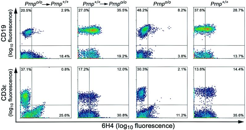
FIG. 2.
Flow cytometric analysis of PrPc expression on PBLs. Reconstitution resulted consistently in CD19-positive B and CD3ɛ-positive T lymphocytes of graft origin, as confirmed by the expression of PrPc and detected by 6H4 antibody staining. Numbers indicate the percentage of cells in the quadrants. Analyses were done on WBCs 6 to 8 weeks after the grafting procedure described. Ordinate, logarithm of fluorescence intensity for B cells (CD19) or T cells (CD3ɛ); abscissa, logarithm of fluorescence intensity for PrP (6H4).

