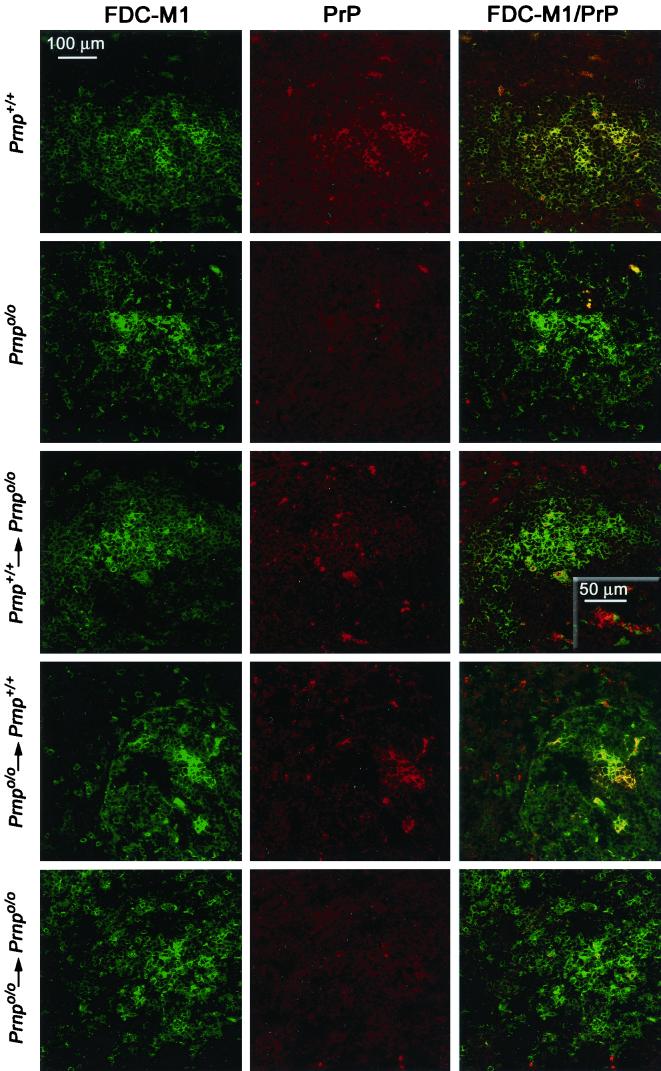
FIG. 3.
Confocal double-color immunofluorescence analysis of splenic germinal centers of reconstituted and inoculated mice and noninoculated controls. Thirty-four days after i.p. scrapie challenge, spleen sections of fetal liver chimeras were stained with FDC-M1 antibody to FDCs (green color, left column) and XN polyclonal antiserum to PrP (red color, middle column). Noninoculated Prnp+/+ ([129Sv × C57BL/6]F1) and Prnpo/o ([129Sv × C57BL/6]Fx) mice served as controls. In spleens of noninoculated wild-type mice or prion-infected wild-type mice grafted with Prnpo/o FLCs (Prnpo/o→Prnp+/+), the signal for PrP is confined to FDC-M1-positive areas (yellow color, first and fourth rows). Instead, Prnpo/o mice reconstituted with Prnp+/+ FLCs (Prnp+/+→Prnpo/o) show PrP-positive cells both in FDC-M1-positive areas (yellow color, third row) and in FDC-M1-negative compartments surrounding the follicular dendritic network (red color, third row).

