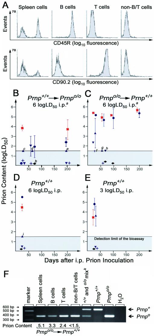
FIG. 4.
Distribution of prions in spleens of scrapie-infected mice. (A) FACS analysis of MACS-purified spleen cell fractions originating from scrapie-infected mice. MACS sorting resulted in >97% of the sorted cell type; therefore cross-contamination could be excluded to a large extent. Anti-CD45R (B220) antibodies were used to label B cells, and anti-CD90.2 (Thy 1.2) was used for T cells. Gating for lymphocytes by forward and side scattering did not interfere with the readout. (B to E) Infectivity levels in splenic fractions and WBCs of FLC-reconstituted and control mice. Fractions were transmitted i.c. in groups of three to four tga20 indicator mice. Symbols: blue circles, spleen cells; blue triangles, B cells; blue inverted triangles, T cells; blue crosses, non-B/T cells; red crosses, stromal fraction; black rhombi, WBCs. Within-group standard deviations are indicated only if they exceed ±0.15 log LD50. Infectivity contents relate to each cellular fraction of individual spleens. (Footnotes) a, similar groups of cell subsets were transmitted after primary inoculation with 3 log LD50; all these fractions were devoid of prions. b, in each of the two separate transmissions of the stromal fraction of this group, all four indicator animals died of intercurrent death within 72 h after i.c. administration, most likely due to brain emboli or toxic contaminations of this homogenate. If one or more animals survived >180 days, the titer was assumed to be close to the detection threshold of the bioassay. For these samples (labeled with small italic letters), the number of animals succumbing to scrapie out of the number of indicator animals inoculated and, in parentheses, incubation time (days) to terminal scrapie in individual mice were as follows: c, one of three (157); d, two of four (110, 116); e, one of four (128); f one of four (115); g, two of four (90, 145); h, two of four (87, 90); i, two of three (90, 93). (F) PCR of spleen cells and splenic B, T, and non-B/T cells. Infectious splenic lymphocytes, derived 34 days after primary inoculation from Prnp+/+ mice reconstituted with Prnp null FLCs, were tested for the presence of the Prnp gene by PCR. Only the Prnpo/o genotype of reconstituting cells was identified in these infectious splenic cell fractions. Prion contents are indicated in log LD50 per fraction of each spleen. k, sample derived from spleen cells known to contain Prnp+/+ and Prnpo/o DNA.

