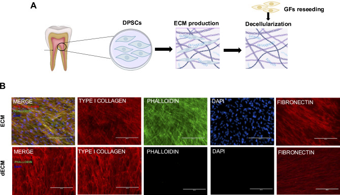
Fig. 1.
Characterization of the extracellular matrix and decellularized extracellular matrix. (A) Conceptual framework depicting the experiment from ECM production to reseeding of gingival fibroblasts (GFs) on decellularized ECM. (B) Both ECM samples exhibited positive staining for TYPE I COLLAGEN and FIBRONECTIN. The intracellular cytoskeleton and nuclei were visualized using phalloidin and DAPI, respectively. No residual cells were observed, as indicated by negative actin and DAPI staining in the dECM

