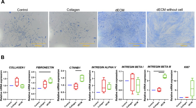
Fig. 5.
The effects of decellularized extracellular matrix derived from dental pulp stem cells (DPSCs) on gingival fibroblast proliferation. (A) Colony-forming unit cell morphology is represented by the colorization of Coomassie blue, while dECM without cell staining was used to distinguish between protein staining and the actual cell-forming colonies. (B) mRNA expression of GFs seeded on dECM at 48 h. Scale bars represent 1000 μm. The data are presented as the means ± standard errors (SEs). Statistical significance is denoted by bars, indicating a significant difference between groups (p < 0.05)

