FIGURE 1.

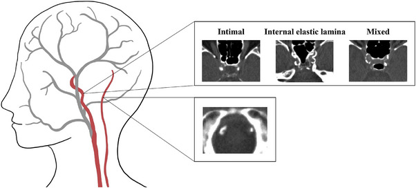
Examples of intracranial internal carotid artery calcification (top) and vertebrobasilar artery calcification (bottom) as seen on non‐contrast computed tomography (CT).

Examples of intracranial internal carotid artery calcification (top) and vertebrobasilar artery calcification (bottom) as seen on non‐contrast computed tomography (CT).