Figure 7.

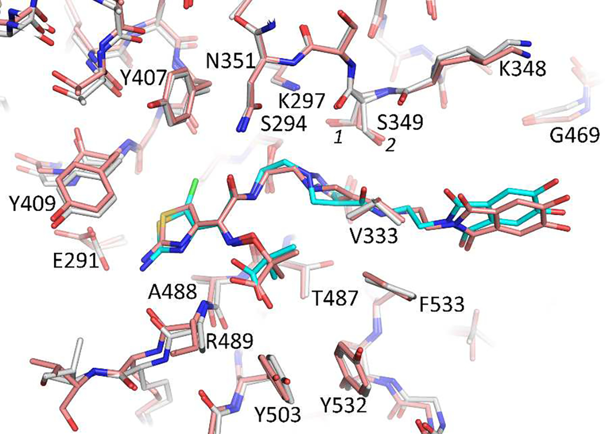
Superpositioning of the 1 and 2 bound structures of P. aeruginosa PBP3. The carbon atoms of the 1:PBP3 complex is shown in salmon color; while 2 is colored cyan with its protein shown in white.

Superpositioning of the 1 and 2 bound structures of P. aeruginosa PBP3. The carbon atoms of the 1:PBP3 complex is shown in salmon color; while 2 is colored cyan with its protein shown in white.