. 2024 Jun 21;17(3):304–308. doi: 10.3400/avd.cr.24-00024

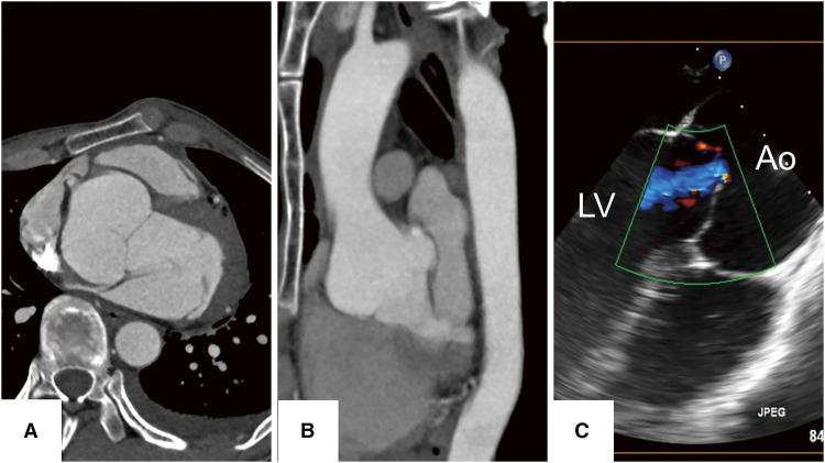
Fig. 1 Findings of preoperative examinations. (A) The axial image of the preoperative CT scan revealed a dilated sinus of Valsalva with a size of 55 mm, which dilated up to 14 mm per 10 years. (B) The sagittal image of CT showed a dilated Valsalva sinus. (C) Echocardiography showed aortic root enlargement and AI. At the end of the diastolic period, a central coaptation defect was observed. AI: aortic valve insufficiency; Ao: aorta; CT: computed tomography; LV: left ventricle.

Fig. 1 Findings of preoperative examinations. (A) The axial image of the preoperative CT scan revealed a dilated sinus of Valsalva with a size of 55 mm, which dilated up to 14 mm per 10 years. (B) The sagittal image of CT showed a dilated Valsalva sinus. (C) Echocardiography showed aortic root enlargement and AI. At the end of the diastolic period, a central coaptation defect was observed. AI: aortic valve insufficiency; Ao: aorta; CT: computed tomography; LV: left ventricle