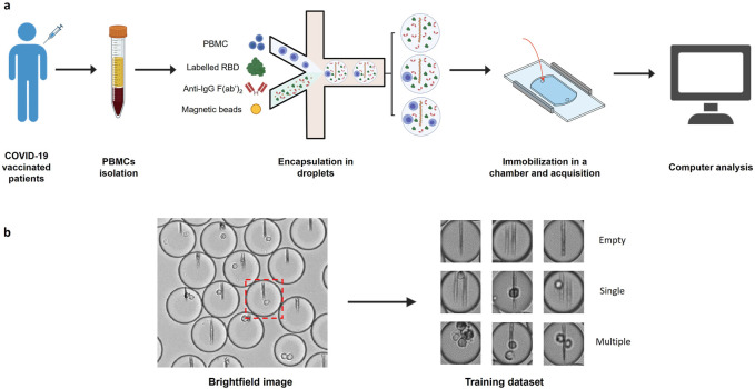
FIGURE 1.
Image datasets construction. (A) PBMCs were isolated from COVID-19 vaccinated patient blood and injected into a microfluidic chip together with a mixture of anti-human-κ chain-VHH-coated paramagnetic nanobeads, fluorescent antigen (recombinant SARS-CoV-2 Spike Receptor Binding Domain) and fluorescent anti-human IgG F(ab’)2. The mix of cells and reagents was encapsulated in droplets using a flow focusing technique. The produced droplets were introduced and immobilized in an observation chamber. A magnetic field generated by two magnets was applied to the chamber so that the nanobeads form a vertical line. (B) Brightfield images of the chamber were acquired with an optical microscope. Images of each droplet were cropped (red square) and manually sorted into 3 populations (empty droplets, single cell droplets, and multiple cell droplets).

