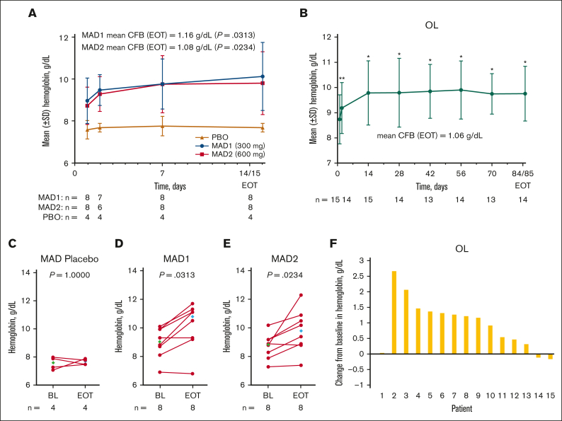
Figure 4.
Change in Hb response in patients with SCD (MAD and OL cohorts). Mean (± SE) Hb concentration over time in the MAD (A) and OL (B) cohorts. Values for mean change from BL at EOT are shown on the graphs (A-B). In the MAD cohorts, EOT was equal to the day 15 value, if available, otherwise EOT was equal to day 14 (A). In the OL cohort, EOT was equal to the day 85 value, if available, otherwise EOT was equal to day 84 (B). Scatterplots at BL and EOT for MAD pooled placebo (C), MAD1 (D), MAD2 (E), and OL (F); each data point corresponds to data from 1 patient. Median BL and EOT values shown in green and blue diamonds, respectively (C-F). Paired BL and EOT data points from each patient are connected by a line. In the MAD cohorts (A), P values were based on Wilcoxon signed rank tests to test the changes at EOT from BL. In the OL cohort (B), Hb values with statistical significance as compared with BL were identified using asterisks (∗P ≤ .0001; ∗∗P < .01) at their scheduled visits, based on MMRM, which included Hb values as a dependent variable, and a fixed effect of scheduled visits during the treatment period, with unstructured covariance matrix to model the within-patient variance-covariance errors. Statistical tests were not performed for the visits after EOT. P value in the scatterplots are from a Wilcoxon matched-pairs signed rank test (C-F). BL, baseline; CFB, change from baseline; EOT, end of treatment; Hb, hemoglobin; MAD, multiple ascending dose; MMRM, mixed model for repeated measurement; OL, open-label; PBO, placebo; SE, standard error; SCD, sickle cell disease; SD, standard deviation.

