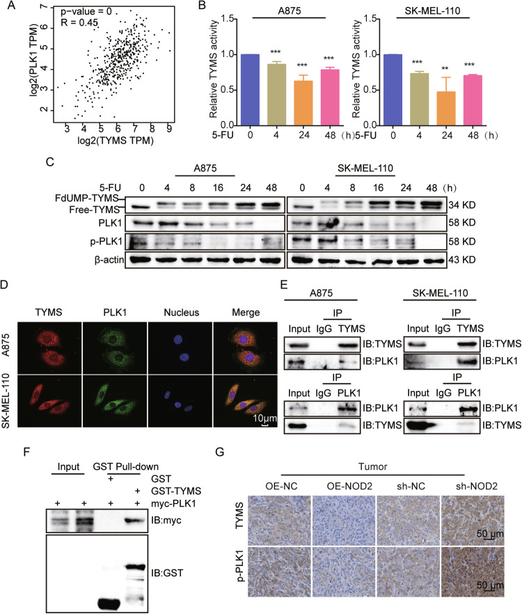
Fig. 8. TYMS regulates PLK1 expression and activation through interaction with PLK1.
A Correlation analysis of TYMS and PLK1 in melanoma in the GEPIA database. B A875 and SK-MEL-110 cells were treated with 5-FU (5 μg/ml) for 0, 4, 24, and 48 h, then evaluated for TYMS activity by ELISA kit. C A875 and SK-MEL-110 cells were treated with 5-FU (5 μg/ml) for 0, 4, 8, 16, 24, and 48 h. Western blot assays to detect the expression of TYMS, PLK1, and p-PLK1. D IF assay to analyze the localization of TYMS and PLK1 in A875 and SK-MEL-110 cells. Scale = 10 μm. E CO-IP assay to detect the interaction between TYMS and PLK1 in A875 and SK-MEL-110 cells. F GST Pull-down assay detects direct interaction between TYMS and PLK1. G IHC staining of TYMS and p-PLK1 in NOD2 overexpression and knockdown tumors. Scale = 50 μm. Data are expressed as the mean ± SD. Student’s t-test and one-way ANOVA were used to compare the differences. **P < 0.01, ***P < 0.001.

