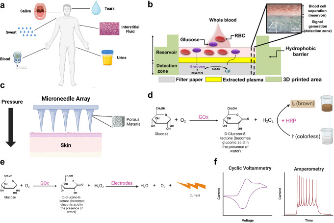
Fig. 1.
Example microfluidic devices and detection methods for glucose testing. a A schematic illustrating bodily fluids, including blood, interstitial fluid, saliva, sweat, tears, and urine, that have been explored for glucose detection. b A paper-based microfluidic blood glucose testing device with colorimetric glucose detection [64]. c Porous microneedle array-driven extraction of interstitial fluid for glucose testing (recreated from [74]). d An example chemical reaction in colorimetric glucose detection with iodide as the chromogenic agent, which is reduced to brown-colored molecular iodine in the presence of H2O2. Other chromogens such as a mixture of 4-aminoantipyrine (AAP) and 3,5-dichloro-2-hydroxybenzenesulfonic acid (DHBS) can be used in place of iodide [80]. e An example electrochemical assay for glucose detection, which generates a detectable current upon the reduction of H2O2. f Cyclic voltammetry and amperometry, two commonly used methods to characterize the electrical current generated by an electrochemical glucose detection system. (a, d, e, f) Created with Biorender. (b) Reproduced from Park C, Kim HR, Kim SK, Jeong IK, Pyun JC, Park S. Three-Dimensional Paper-Based Microfluidic Analytical Devices Integrated with a Plasma Separation Membrane for the Detection of Biomarkers in Whole Blood. ACS Appl Mater Interfaces. 2019;11:36428–36,434 [64]. Copyright permission from ACS Publications (CC License). (c) Recreated from Takeuchi K, Takama N, Kinoshita R, Okitsu T, Kim B. Flexible and porous microneedles of PDMS for continuous glucose monitoring. Biomed Microdevices. 2020;22:79 [74]

