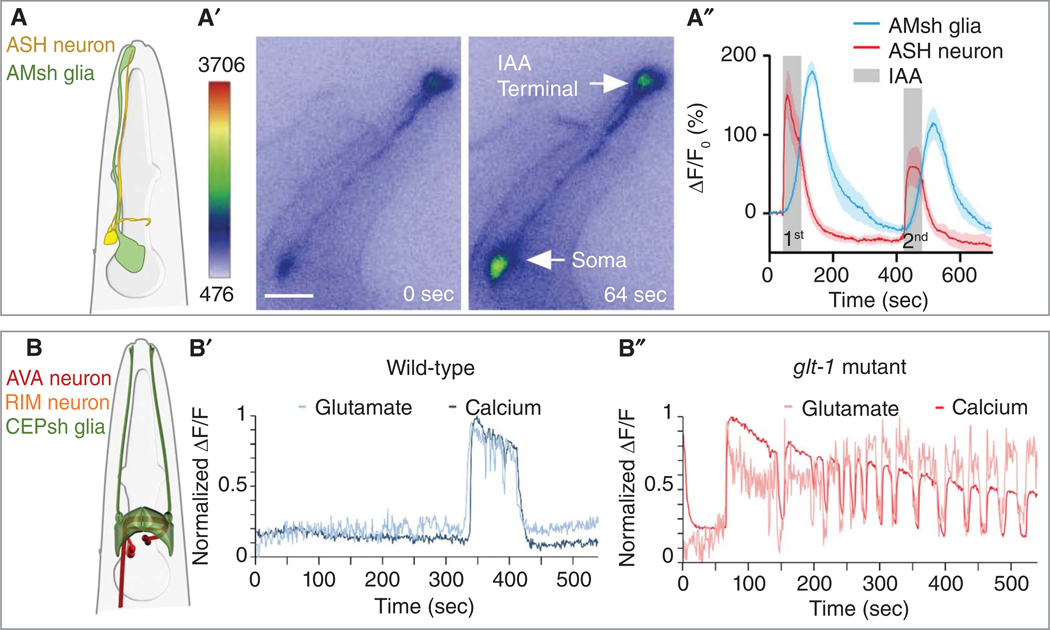
Figure 4.
Caenorhabditis elegans glial functions in animal behavior. (A–A′′) Schematic (A), representative micrograph (A′) (scale bar, 20 μm), and Ca2+ transient quantification (A′′) in AMsh glia and ASH neuron upon two pulses of isoamylalcohol (IAA) stimulation. (B–B′′) Schematic (B) depicting the region where CEPsh posterior processes contact AVA and RIM neurons. Representative traces of spontaneous glutamate (light, measured by iGluSnFR) and calcium (darkline, measured by GCaMP) dynamics near the AVA neuron in wild-type (B′) and glt-1 mutant (B′′) animals. (Images in A are reprinted with permission from Duan et al. 2020. Images in B are reprinted from Katz et al. 2019 under a Creative Commons Attribution 4.0 International License.)

