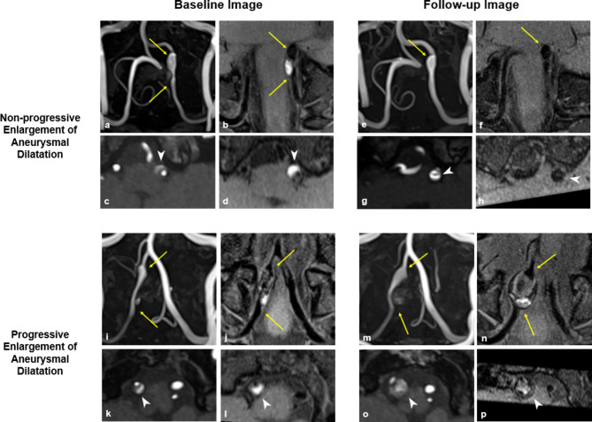
Fig. 3.
Baseline and Follow-up Images in Cases of Non-progressive and Progressive Enlargement of Aneurysmal Dilatation. (a–h) Representative case of non-progressive enlargement of aneurysmal dilatation. (a, b) Baseline images show dissection in the left vertebral artery (arrow) with an increased outer diameter compared to the adjacent normal-looking artery. (c, d) Axial images at the point of maximal outer diameter shows double lumen with dissecting flap and intramural hematoma (arrow head). The remodeling index, normalized wall index, and relative signal intensity of intramural hematoma (rsIMH) were 2.49, 0.59, and 3.09, respectively. (e, f) Follow-up images after 6 months of symptom onset shows residual aneurysmal dilatation of the dissected artery (arrow). (g, h) Axial images demonstrates dissecting flap (arrow head) but no intramural hematoma in both TOF-MRA and VW-MRI. The remodeling index, normalized wall index, and rsIMH were 2.22, 0.64, and 1.33, respectively. (i–p) Representative case of progressive enlargement of aneurysmal dilatation. (i, j) Baseline images show dissection in the right vertebral artery (arrow) with an increased outer diameter. (k, l) Baseline axial images show double lumen with dissecting flap and intramural hematoma (arrow head). The remodeling index, normalized wall index, and rsIMH were 3.43, 0.67, and 3.94, respectively. (m, n) Follow-up images after 6 months of symptom onset show continuous enlargement in the diameter of the dissected artery (arrow). (o, p) Axial images demonstrate the onion-skin appearance of a multilayered hematoma. The remodeling index, normalized wall index, and rsIMH were 26.08, 0.99, and 4.89, respectively.

