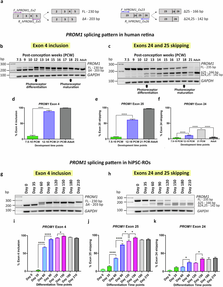
Fig. 1. PROM1 alternative splicing in retina in vivo and in vitro.
a Schematic representation of the primers used to amplify exon 4 (on the left) and exons 24 and 25 (on the right). b–f RT-PCR analysis of PROM1 splicing during human retinal development: b Exon 4 PROM1 and c Exon 25 PROM1 splicing pattern; d–f Quantification of exon 4 inclusion, exon 25 skipping and exon 24 skipping. Data are shown as mean ± SEM (n = 3–7). Statistical significance was assessed using one-way ANOVA with Sídák’s post hoc test. ****p < 0.0001. g–k RT-PCR analysis of PROM1 splicing during hiPSC-ROs differentiation: g Exon 4 PROM1 and h Exon 25 PROM1 splicing pattern; i–k Quantification of exon 4 inclusion, exon 25 skipping and exon 24 skipping. Data are shown as mean ± SEM (n = 3). Statistical significance was assessed using one-way ANOVA with Sídák’s post hoc test. *p < 0.05, ****p < 0.0001. FL Full-length product, △4 Exon 4 skipped, △25 Exon 25 skipped, △24,25 Exons 24 and 25 skipped, PCW Post-conception week.

