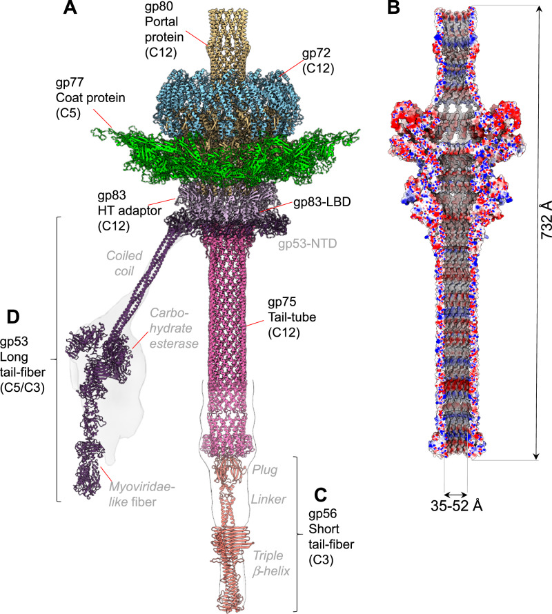
Fig. 2. DEV tail apparatus.
A Composite ribbon diagram of DEV tail reconstructed from FF virions. Tail factors identified de novo in the C12 localized reconstruction include the portal protein gp80 (yellow), the ejection protein gp72 (blue), the HT-adapter gp83 (light purple), and the tail tube gp75 (magenta). B Cross section of an electrostatic surface representation of the DEV tail channel. Red, blue, and white represent negative, positive, and neutral charges near the surface. C–D AlphaFold models for the short-tail fiber gp56 and long-tail fiber gp53 overlaid to low-resolution localized reconstructions shown as semitransparent surfaces. Individual tail factors are color-coded, as in panel (A).

