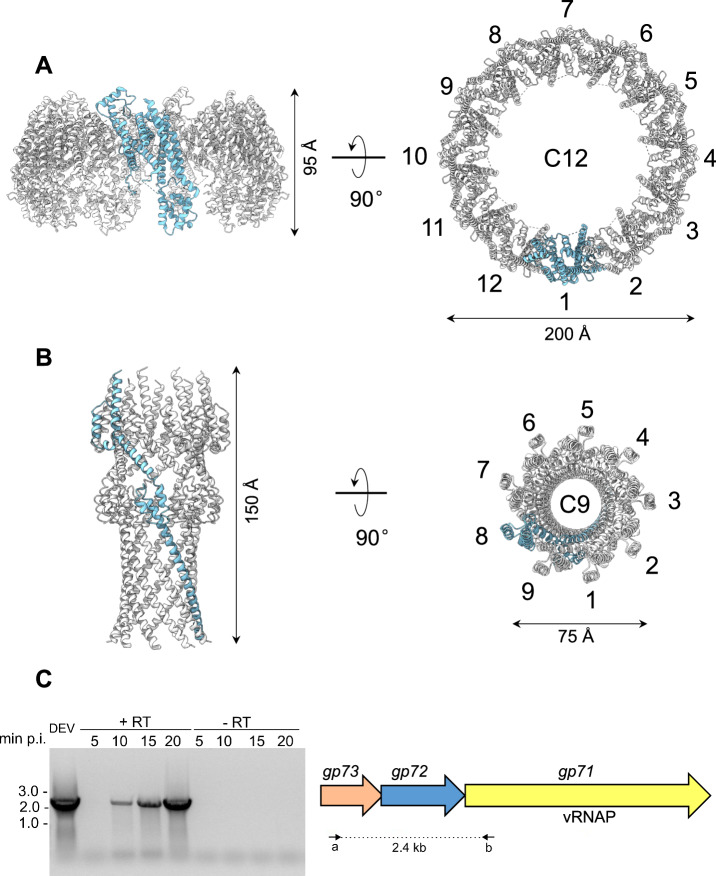
Fig. 5. Quaternary structures of DEV ejection protein gp72 pre- and post-ejection.
A The quaternary structure of DEV gp72 from FF virions determined in situ. Twelve gp72 subunits surround the portal protein, generating a ~ 200 Å wide ring. B Cryo-EM structure of the recombinant nonameric gp72 determined at 3.65 Å resolution in the post-ejection conformation. In panels (A and B) only one protomer is colored in cyan, whereas all other subunits are light gray. (C) DEV gp71, gp72, and gp73 genes are co-transcribed as an operon. (Left panel) Agarose gel electrophoresis of RT-PCR products. RNA samples extracted from PAO1 cultures at different time points post-infection (p.i.) with DEV (e.g., 5, 10, 15, 20 minutes) were reverse-transcribed ( + RT) or not (negative control, –RT) and used as templates for PCR amplification. Migration of MW (kb) markers is shown on the left. The assay was repeated three times with similar result. (Right panel) Schematic diagram of DEV ORFs encoding gp71, gp72, and gp73. Arrows represent the position of oligonucleotides used for amplification, yielding a 2.4 kb long amplification product. Source data are provided as a Source Data file.

