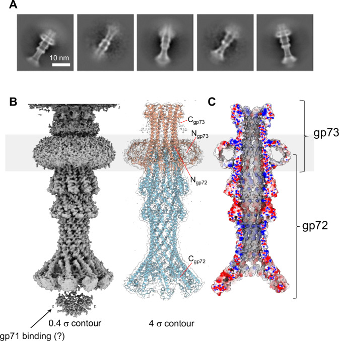
Fig. 6. DEV ejection proteins gp72 and gp73 form a tube-shaped complex.
A Representative 2D class averages of the gp72:gp73 complex. B 3D reconstruction of the gp72:gp73 complex visualized at low (left) and high (right) contours. The atomic models of gp73 and gp72 are overlaid to a semitransparent density calculated at 3.15 Å resolution. In gray is the putative position of the bacterium’s outer membrane. C The cross-section of an electrostatic surface representation of gp72:gp73 shows the lumen and surface charge inside the channel. Red, blue, and white represent negative, positive, and neutral charges near the surface.

