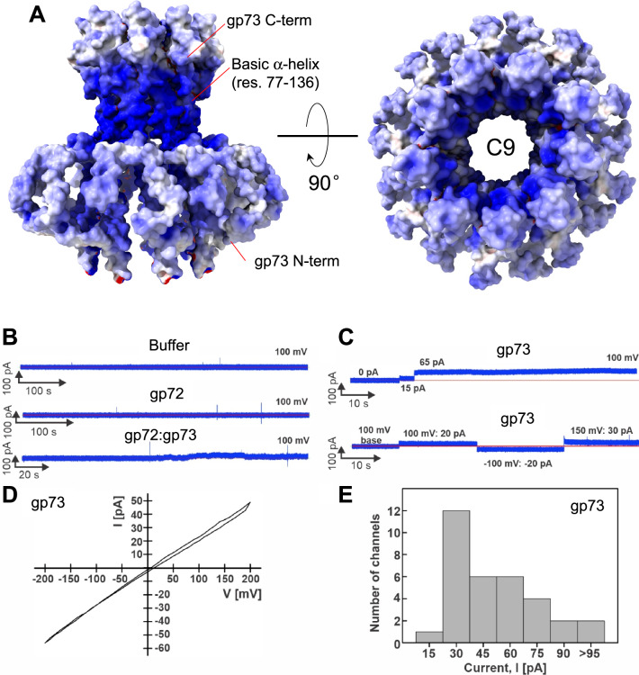
Fig. 7. Lipid bilayer experiments with purified DEV ejection protein gp73.
A The electrostatic surface representation of nonameric gp73 reveals a significant positive charge, mainly in the α-helical core. B,C Lipid bilayer experiments were performed at 100 mV applied potential in diphytanoylphosphotidylcholine (DPhPC) membranes bathed in 1 M KCl, 10 mM HEPES, pH 7.4 electrolyte. The protein samples were added to the grounded trans side of the cuvette, which had 100 µm SU-8 aperture. B Representative current traces. Top: 15 µl protein buffer in the cuvette. Six membranes were recorded with 1 – 15 µl of the protein buffer, and no activity of the buffer was observed. Middle: gp72 current trace. Seven membranes with up to 24 µg of gp72 in the cuvette were recorded, and no channel activity was observed. Bottom: gp72:gp73 complex. 19 membranes were recorded, and only one shown here had 10 – 20 pA fluctuations around the baseline when 10 µg of protein sample was in the cuvette. C Representative current traces of gp73. Top: Two insertions of gp73 (750 ng protein in the cuvette) with amplitudes of 15 pA, and 65 pA. Bottom: Continuous current trace of a single gp73 insertion (900 ng in the cuvette) at indicated voltages. D Current-voltage curve of one gp73 pore inserted in the DPhPC membrane at a voltage range of −200 to 200 mV. E Histogram of single-channel current amplitudes of gp73 at 100 mV. A total of 33 channels were observed with a mode current of 30 pA.

