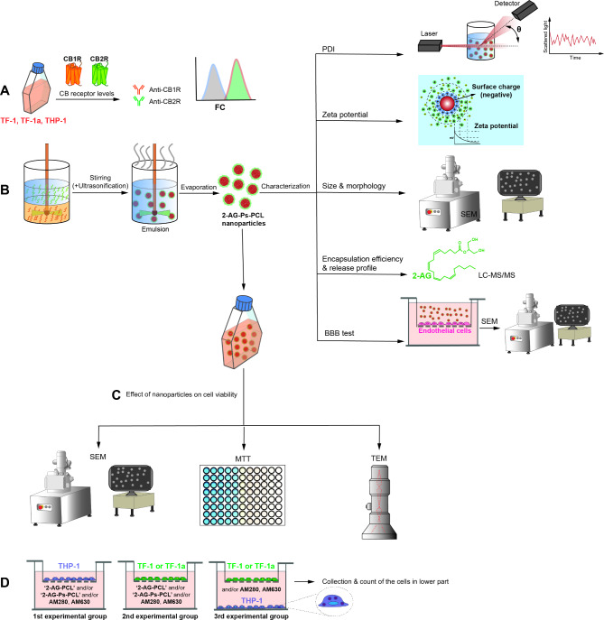
Fig. 1.
Schematic design of the study. A We detected CB1R and CB2R expression in TF-1, TF-1a and THP-1 cells. B We prepared the nanoparticles using the emulsion evaporation method and characterized them by analyzing their PDI, zeta potential, surface charge, size, morphology, EE, release profile and invasion profile using DLS, SEM, LC‒MS/MS and in vitro invasion assays, respectively. C We analyzed the effect of nanoparticles on TF-1, TF-1a and THP-1 cell viability using SEM, TEM and MTT assays. D We detected the migratory effect of nanoparticles on TF-1, TF-1a and THP-1 cells using a transwell migration assay

