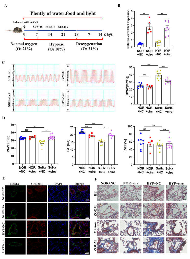
Fig. 7.
Over-expression of circSSR1 in vivo inhibit the symptoms of pulmonary hypertension. A, Construction of hypoxia model mouse. B, Specificity over-expression circSSR1 in the mouse lung tissue (n = 5–8). C, Right ventricular (RV) systolic pressure (RVSP), RV/left ventricular (LV) + S weight ratio and heart rate in the HYP-PH mice models. D, Pulmonary arterial velocity time integral (PAVTI) and Pulmonary artery acceleration time (PAT), left ventricular ejection fraction (LVEF) in the NOR + AAV5–negative control (NC), NOR + AAV5, HYP + AAV5–negative control (NC), and HYP + AAV5 groups are shown (n = 5–10). E, Immunofluorescence of GSDMD in mouse lung tissue section. Scale bars, 50 μm. F, HE and Masson staining showed circSSR1 over-expression prevented and reversed wall thickening. Scale bars, 100 μm. All values are presented as the mean ± SEM. *p<0.05, **p<0.01, ***p<0.001. NOR, normoxia and HYP, hypoxia. NC: negative control

